
The Scleral Lens Education Society: Case Report 2
Dirk J Booysen
Dip. Optom FOA(SA), MCOptom(UK), TMOD(PCO), CAS(NECO), DOptom(Aston)
248 Voortrekker Road
Monument
Krugersdorp
Republic of South Africa
Abstract
Keratoconus is a bilateral asymmetric non-inflammatory corneal degeneration characterised by localised corneal thinning which leads to ectasia or protrusion of the thinned cornea causing high myopia and irregular astigmatism that affects the visual acuity and the patient’s quality of life. Pellucid marginal degeneration is a rare, bilateral progressive, non-inflammatory corneal ectasia characterized by thinning of the inferior peripheral cornea leading to high against-the-rule or irregular astigmatism. Both keratoconus and pellucid marginal degeneration are classified as “ectatic corneal diseases”. The most important objectives of non-surgical treatment is firstly to halt progression and the secondly to provide visual rehabilitation. Contact lenses are extremely beneficial to correct vision in many patients but does not slow or halt progression of ectasia.
This case report deals with fitting rigid gas permeable corneal and rigid gas permeable mini-scleral lenses to visually rehabilitate a patient with pellucid marginal degeneration and keratoconus.
Key Words
Keratoconus, scleral lenses, corneal ectasia, hydrops, pellucid marginal degeneration, and rigid gas permeable lenses
Introduction
Keratoconus is a bilateral asymmetric non-inflammatory corneal degeneration characterised by localised corneal thinning which leads to ectasia or protrusion of the thinned cornea 1,2. The corneal thinning and protrusion can occur anywhere on the cornea but is most commonly found in the inferior and central cornea and this leads to high myopia and irregular astigmatism that affects the visual acuity and the patients quality of life 1. Keratoconus, pellucid marginal degeneration, keratoglobus and progressive iatrogenic ectasia is classified under “ectatic corneal diseases” while conditions such as Terriens marginal degeneration, dellen and inflammatory melts is not classified as ectatic diseases but “thinning disorders” of the cornea 2. Keratoconus typically presents during the second decade of life during puberty and progresses until the forth decade when it stabilizes 1. Histopathologically , there are three signs that typically characterise keratoconus; corneal thinning, Bowman’s layer breaks, and iron deposits within the corneal epithelium’s basal layer (Fleischer’s rings) 1. The epithelial basal cells degenerate and decrease in density and grow toward Bowman’s layer which often show breaks filled with collagen from the stroma. In the stroma the number of lamellae, keratocytes are decreased leading to changes in the gross organization of the anterior stroma lamellae and collagen fibrils particularly around the apex of the cone 1. The corneal nerves have thicker bundles, reduced density and sub-epithelial plexuses compared to normal corneas 1. Descemet’s membrane and the endothelium are usually unaffected until the late stages of the disease when breaks can occur leading to disruption of the endothelial pump mechanism which results in corneal hydrops 1. Despite intensive research into the aetiology and pathogenesis of keratoconus, the causes and mechanisms for its development are poorly understood and there is no primary pathophysiologic explanation for keratoconus 2. However, several hypotheses have been proposed which include genetic, environmental, biochemical, mechanical and biomechanical mechanisms. Furthermore keratoconus associations with other diseases are well documented 1,2.
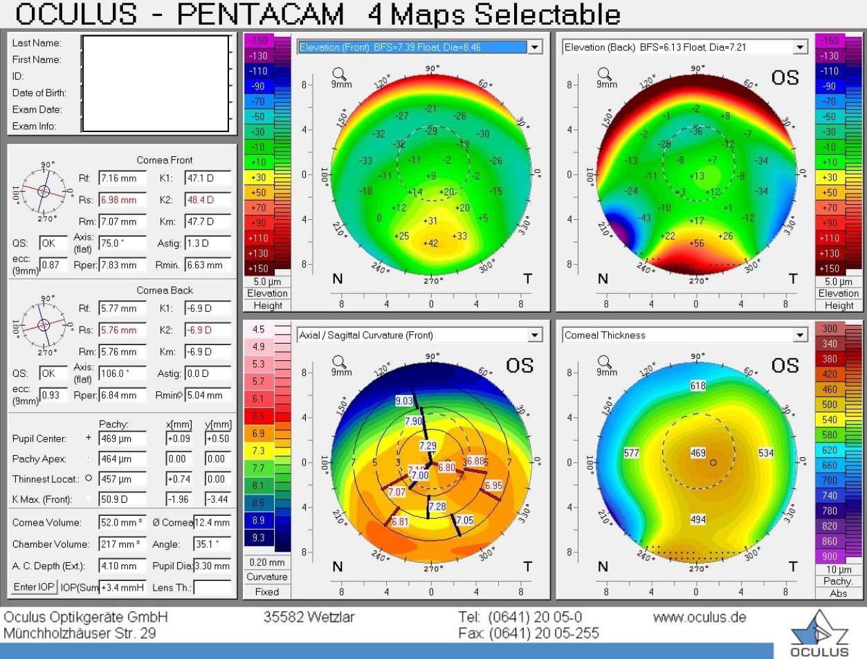
Pellucid marginal degeneration (PMD) is a rare, bilateral progressive, non-inflammatory corneal ectasia characterized by thinning of the inferior peripheral cornea 3-6. The area of protrusion typically occurs above the area of thinning and not within the area of thinning as seen with keratoconus. Topography maps of the cornea show inferior corneal steepening with a “butterfly” pattern and high against-the rule-astigmatism (Figure 7). Like keratoconus PMD is considered an “ectatic corneal disease” and patients present with symptoms slightly later in life, typically in the third to fifth decade of life 3-6. PMD is more prevalent in males than females 3 and their seems to be no connection between ethnicity and prevalence 6. Although data regarding the genetic component of PMD is lacking, some articles suggest that it has a similar inheritance pattern to keratoconus 7. PMD progression decreases dramatically with age and the condition tends to stabilize in the fifth to sixth decade. Complications of PMD are similar to those of keratoconus and include corneal hydrops and corneal perforation at or near the area of thinning 3.
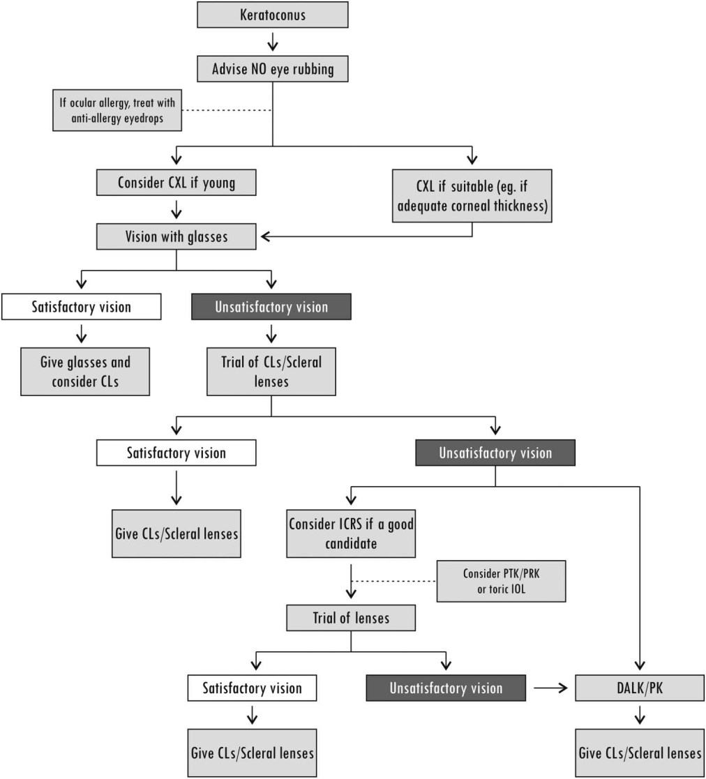
Keratoconus can be managed non-surgically or surgically depending on the severity of the disease. Even in the case of corneal hydrops, non-surgical management should be attempted before keratoplasty 2. The most important objectives of non-surgical treatment is firstly to halt progression and the secondly to provide visual rehabilitation. Among the non-surgical treatment measures verbal guidance regarding the importance of not rubbing one’s eyes and effective treatment of ocular allergy with topical multiple-action anti-allergic medication are probably the most important to halt progression of the disease 1,2. Contact lenses are extremely beneficial to correct vision in many patients but does not slow or halt progression of ectasia 1,2. The flowchart from Gomes et al, 2015 explains the treatment options available to the keratoconus patient (Figure 1).
Management of PMD is similar to that of keratoconus and depends on the severity of the condition. In the early stages of the disease, PMD can be managed with spectacles or soft contact lenses 8. Patients with higher amounts of irregular astigmatism may need rigid gas permeable (RGP) or hybrid lenses to achieve acceptable visual outcomes 8. Fitting conventional corneal RGP lenses is difficult due to the inferior position of the ectasia and aspheric larger diameter RGP’s are required to improve centration, comfort and visual outcomes. Scleral and semi-scleral lenses are viable options in the management of PMD because of the improved centration and apical clearance which limits mechanical irritation of the ectatic cornea 9,10. Studies have shown that visual outcomes are often improved compared to corneal RGP lens designs 9,10. As with keratoconus verbal guidance regarding the importance of not rubbing one’s eyes and effective treatment of ocular allergy with topical multiple-action anti-allergic medication is important in the treatment of PMD 1,2
Case Report
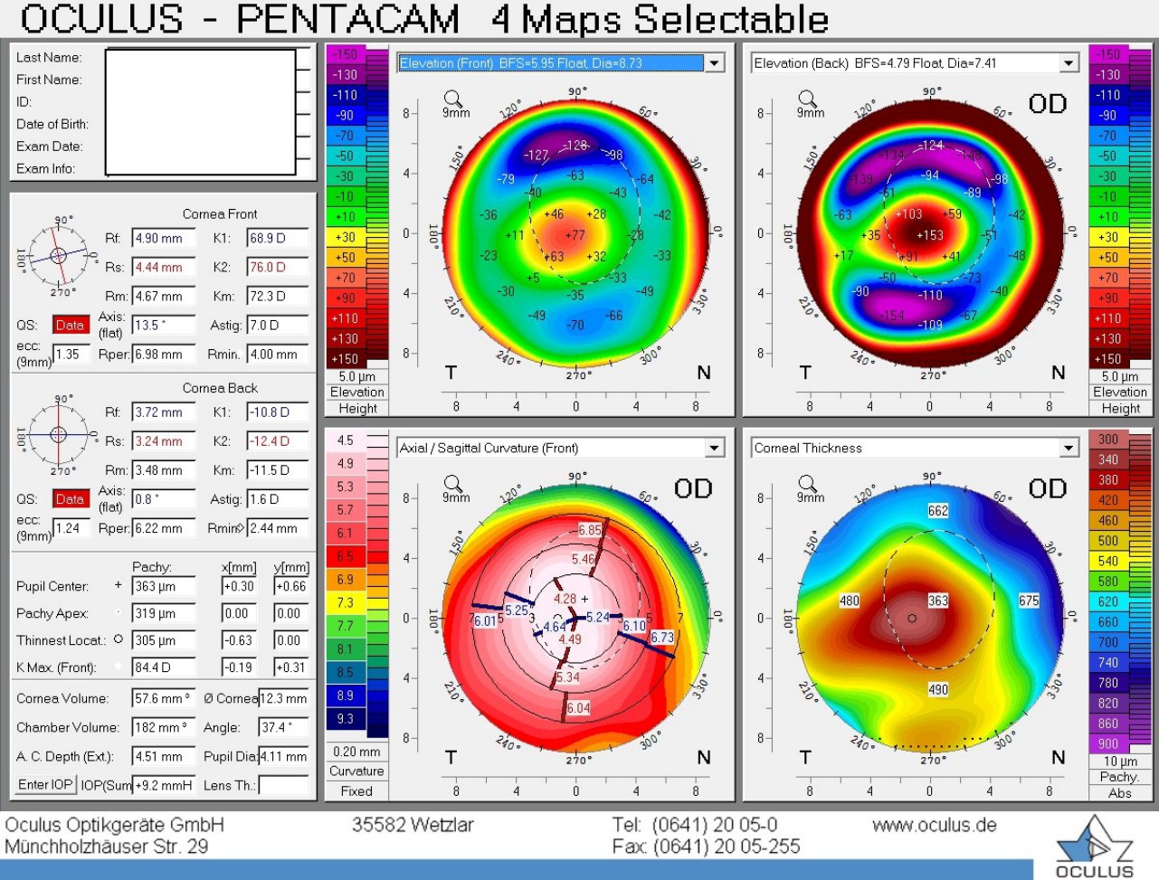
This 35 year old African male patient was refered by a colleugue for contact lens evaluation. On the 30th March 2015 he reported that he had keratoconus and uses a piggy back system for correction of the left eye only. He previously wore an RGP lens on the right eye but apparently suffered a hydrops which resulted in a central scar affecting his comfort and acuity with the RGP lens. At this vist the patient had right finger counting at 2 meters, and left 20/20 acuity with the piggy back system. The RGP in the left eye was as follows: Base curve 6.60/ power -10.00DS/ diameter 10.00mm unknown design with 4 curves. The soft lens used was -0.50/8.50/14.00 Acuvue Oasys. Figures 4 – 7 show the results of my examination in more detail.
Examination and contact lens fitting
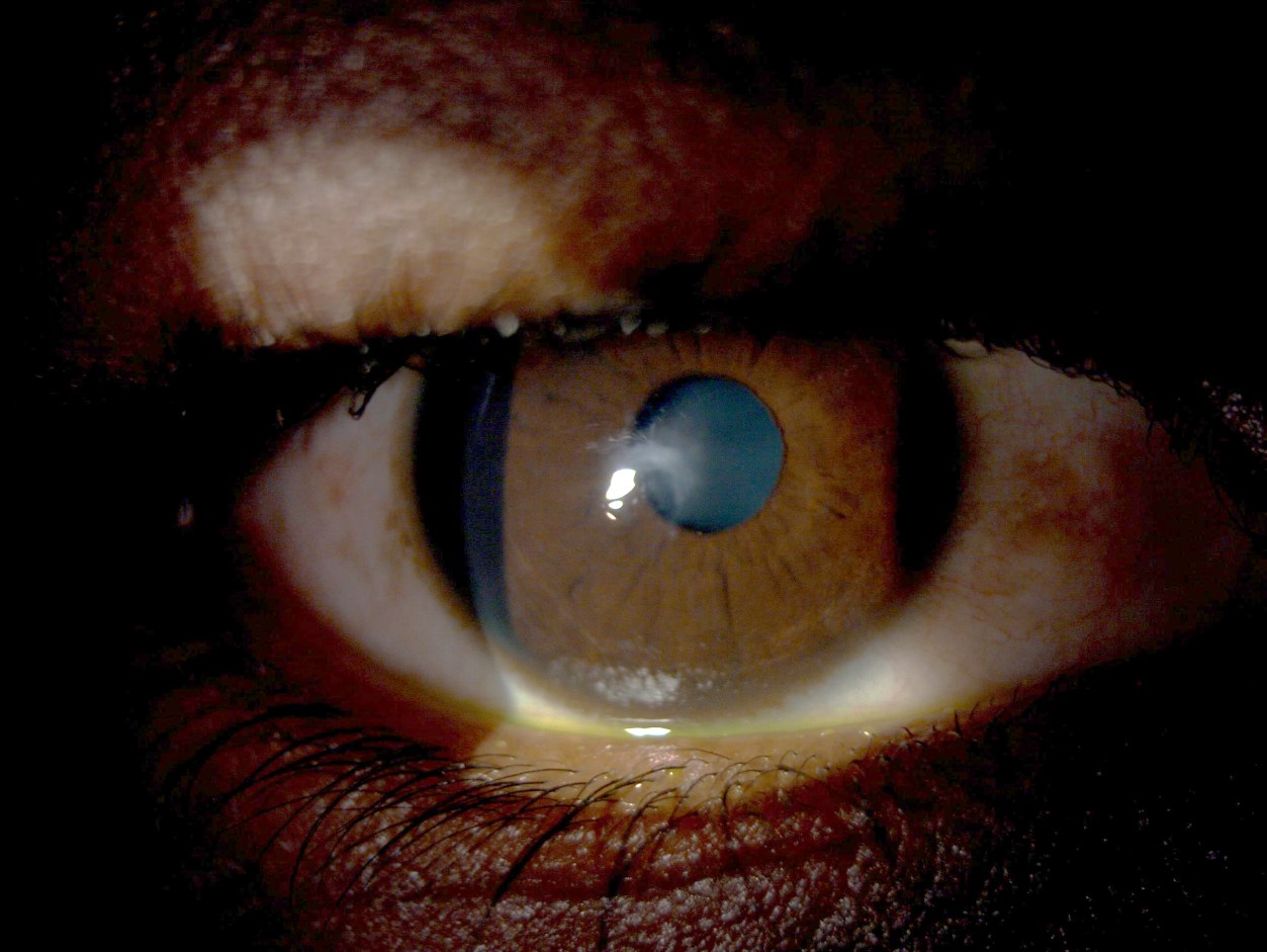
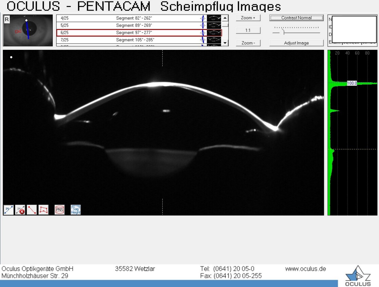
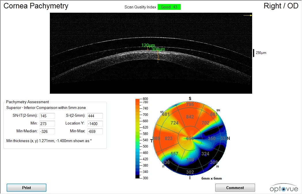
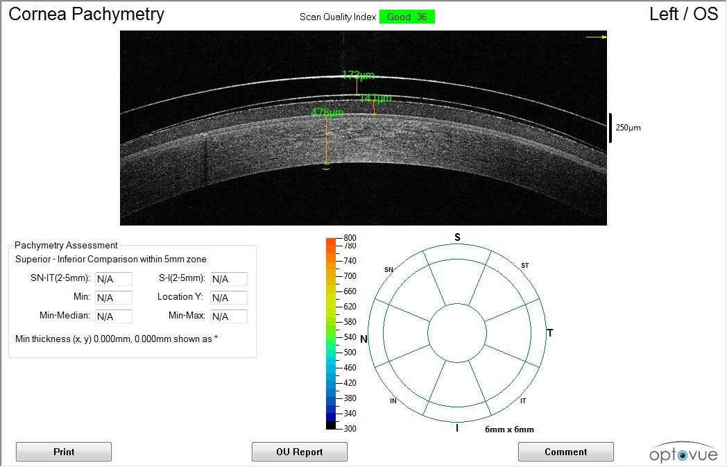
Slitlamp examination of the eyes showed the presence of a central scar in the right cornea extending anterior to posterior around 100 microns deep. Neither cornea stained with NAFL and TBUT’s were 10 and 12 seconds right and left eye respectively. Pentacam pachymetry was 305 microns which corelate well with OCT pachymetry of 304 microns. Front corneal curvature of the righe eye was 68.9D/76.0D with an average of 72.3D and the exterior (from epithelium) anterior chamber depth was 4.51mm. Pachymetry in the left eye was 457 microns and the scan indicates that the patient has moderate PMD in this eye. Front corneal curvature of the left eye was 47.1D/48.4D with and average of 47.7D. The external anterior chamber depth was 4.10mm. Corrected GAT intraocular pressure was right 7+9.2= 16.2mmHg and left 12+3.4=15.4mmHg (Dresden formula). The posterior segments of both eyes were normal with mydriatic examination and cup to disc ratios were 0.4 in both eyes. Both disc were tilted. The patient was phacic and both lenses were clear. Refractive error and vision without lenses was:
R. -6.00/-6.50×20 20/400-
L. -7.00/-3.00×80 20/100-
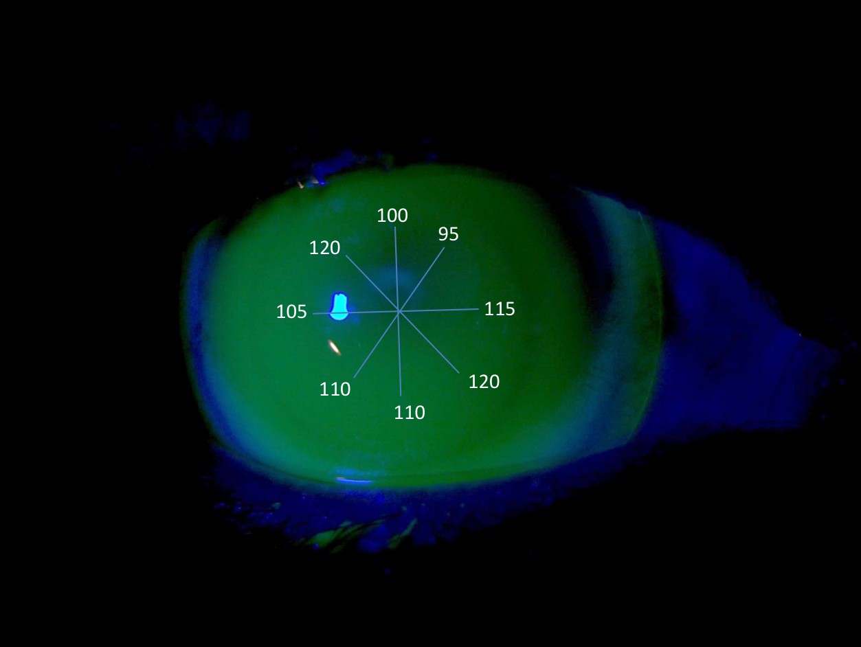
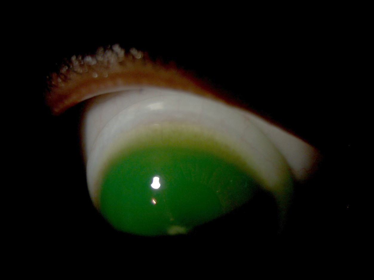
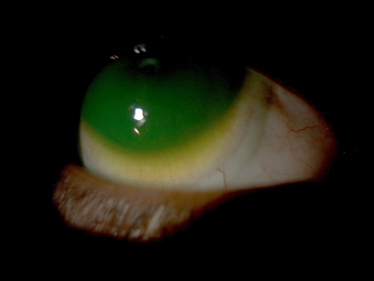
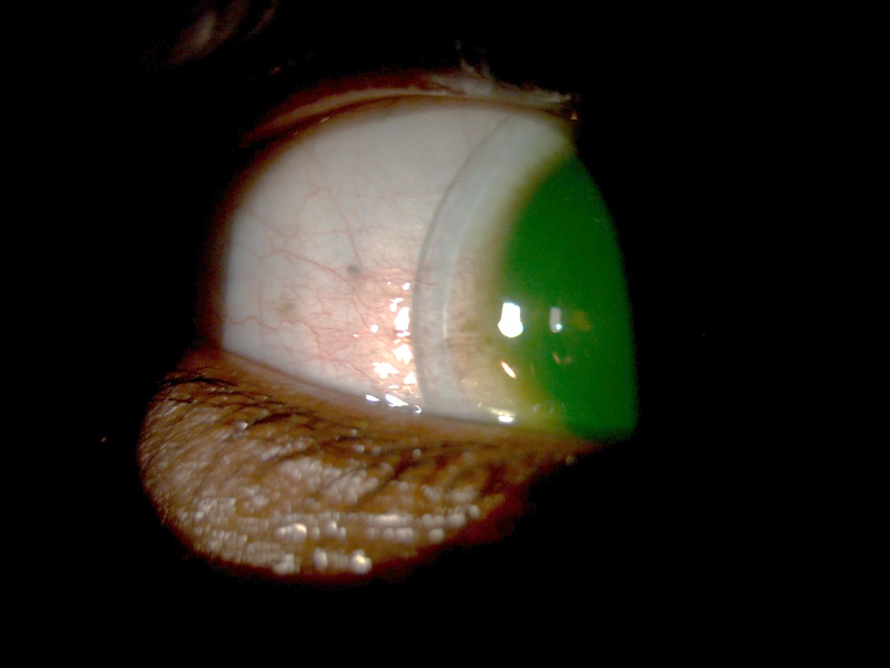
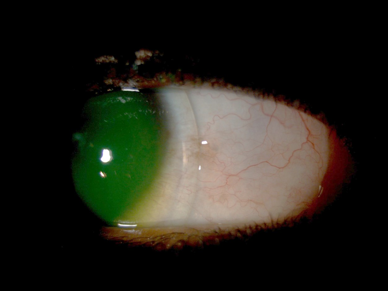
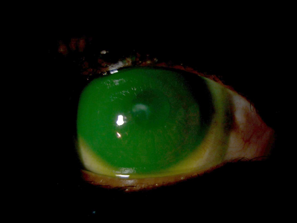
Van Der Worp, 2015 classifies a mini-scleral lens as having a diameter of 15 to 18.00 mm bearing mainly on the sclera (Figure 18) 10. This classification is supported by the Scleral Lens Education Society Nomenclature which defines a mini-scleral lens as a lens that rests entirely on the sclera and is less than 6 mm larger than the horizontal visible iris diameter 11. A mini-scleral lens was selected for the right eye based on the anterior chamber depth + 0.3mm. This shows that the sag of the trial lens should be 4.81mm. Based on this calculation the following lens (Figure 2) was placed on the eye and the accompanying photographs indicate the fluorescein patterns and edge profiles (Figures 8 – 13). Anterior segment OCT was also performed and show corneal clearance (tear lens) of at least 100 microns in all meridians as well as adequate limbal clearance (Figure 14). Over refraction of the plano trial lens was -6.00DS and acuity was 20/80. Althoug autorefraction showed -3.00DCx60 this was rejected with subjective testing and made no difference to the acuity. The final lens ordered was different than the trial lens with a larger diameter, flatter and wider intermediate as well as peripheral curves and deeper sagital depth (Figure 3).
From my obsevation of the current left RGP fit it was evident that the lens fit was too steep and this was rectified by changing the base curve to 6.9mm. The final lens ordered was a Keratocon design with the following paramenters. Base curve 6.9mm/OZD 7.0mm/2nd 7.50mm/8.10mm/3rd 8.10mm/8.70mm/PC 12.25mm/power-8.50/diameter 9.5mm in Boston XO2 (Dk 141) material. Acuvue lenses are no longer availible in South Africa (Johnson & Johnson Vision care withdrew all products and closed franchise on the 6th March 2015) nessesitating a refit with a CooperVision silcone hydrogel lens in the left eye. Parameters of this lens was +0.50/8.40 base curve/diameter 14.20mm/Dk 100/ water content 62%/Avaira/ material Enfilcon A. The lenses were dispensed on the 16th April 2015.
He was instructed on different lens insertion and removal techniques and felt most comfortable using a DMV plunger to remove and insert the lenses. The lens bowl was filled with unpreserved saline (Alcon Polyrinse®) before insertion. Hydrogen peroxide was recommended as a disinfection system (AO Sept) after cleaning and rinsing the lenses with a surfactant (Crystal Cleaner– alcohol based surfactant) and saline respectively. Alcohol based surfactant cleaners are very effective in removing surface lipid and protein deposits with manual rubbing. In combination with hydrogen peroxide sterilization this cleaning regime is very effective for all types of contact lenses significantly simplifying his care regimen. Hydrogen peroxide provides excellent antimicrobial and antifungal efficacy. It works by penetrating the lens material and cleans by expanding the lens matrix and oxidizing microbes. Because of its hypotonic nature and pH of 4.00, H2O2 is also able to break protein and lipid bonds, remove trapped debris and penetrate bacterial biofilms12. H2O2 is highly effective against all microorganisms when used in a 3% concentration but is non-selective in its activity. A two-step 3% hydrogen peroxide solution is also 99.9% cysticidal providing contact times of at least four hours are employed 13.
Follow-up
At follow up examination on the 16th May 2015 the patient reported good comfort and wearing time of the mini-scleral lens as well as the new piggy back system on his left eye. His visual acuity was right 20/40 and left 20/20 which was a signifcant improvement compared to his previous acuity. The corneas were clear with no NAFL staining and he had no ocular inflammation. Corneal clearance remained at 120 microns with the mini-scleral lens. Follow-up visits were scheduled for every 3 months. The last follow-up examination took place on the 10th September 2016. The patient reported no adverse reactions and mainly came in to purchase disposable soft lenses for his left eye. Examination of the mini-scleral lens showed a small chip in the edge which did not cause any ocular problems. The corneas were clear without NAFL staining and vision remained right 20/40 and left 20/20 with the lenses. Corneal clearance with the anterior segment OCT was 110 microns and limbal clearance ±200 microns. The mini-scleral lens was sent to the laboratory for re-edging resulting in a slight decrease in overall diameter to 15.90mm. This did not alter the fit significantly and the lens was dispensed on the 15th September 2015. The next follow-up visit was scheduled for February 2016. I discussed the option of a mini-scleral lens (instead of the piggy-back system) for the left eye with the patient and he will consider this before seeing me again in 2016.
Differential Diagnosis
- Pellucid marginal degeneration (PMD), right eye: Bilateral asymmetrical corneal thinning in the inferior periphery with protrusion superior to the band of thinning. PMD can co-exist with keratoconus and keratoglobus. Corneal topography show a “butterfly” pattern with severe astigmatism and diffuse steepening in the inferior cornea 4,5,14
- Keratoglobus, both eyes: Rare congenital, non-progressive disorder with a circular uniformly thinned cornea. Onset is at birth. Maximal thinning occurs in the mid-periphery of the cornea with protrusion central to the area of maximal thinning 4,5,14
- Iatrogenic ectasia, both eyes: After lamellar refractive surgery such as Laser in situ keratomileusis (LASIK), and rarely surface ablation (PRK), an ectasia similar to keratoconus can develop. Treatment is similar to that of keratoconus 4,5,14. He had no previous eye surgery.
- Posterior Keratoconus, both eyes: Uncommon, sporadic unilateral, non-progressive increase in the curvature of the posterior corneal surface. The anterior surface is normal and visual acuity may be unimpaired due to the similar refractive indices of the cornea and aqueous humour. Two types are recognized; Generalis, where the entire posterior surface curvature increases and Conscriptus, where the increase in curvature is localized either centrally or paracentrally 4,5,14
- Terriens marginal degeneration, dellen and inflammatory melts are not classified as ectatic diseases but “thinning disorders” of the cornea 2 and can be included in the differential diagnosis of PMD of the left eye.
Discussion
Pellucid marginal degeneration has features similar to those seen in keratoconus. This includes progressive thinning and ectasia, hydrops and scarring. However, it differs from keratoconus in the more peripheral location of the thinning, lack of Fleischer’s rings, Vogt striae, a conical shape, and the slightly older age of affected patients 15. Some eye care professionals feel that keratoconus and PMD are different presentations of the same disease 5,7,15,16. Further research of the biomechanical changes and underlying disease processes will reveal if this is the case or not 15. This specific case seems to support the hypotheses that the two diseases are in fact one and the same. Pentacam scans (Figures 6 and 7) clearly show the differences between the two eyes, right showing a central location of the ectasia and left the typical “butterfly pattern” normally associated with PMD. Management of PMD and keratoconus are essentially the same (Figure 1). However, RGP contact lens fitting in PMD can often be difficult due to the inferior location of the ectasia 9. This patient was using a “piggy back” RGP and soft disposable combination on the left eye with good success. He was happy to continue with this fitting modality and specifically wanted a contact lens option for his right eye. As discussed previously a semi-scleral lens was fitted with good results and in future a similar lens will be fitted in the left eye if the patient agrees with that. Surgical correction of PMD is more difficult than in keractoconus and requires large, inferiorly centred grafts which are prone to neovascularization, rejection, and high postoperative astigmatism15.
Oxygen delivery to the cornea is impacted by the scleral lens and the tear lens thickness, especially if the scleral lens was thicker than 350 microns and the tear layer thickness exceeds 200 microns 17. In order to minimize hypoxia induced swelling with scleral lenses, lenses with high Dk values (>150), maximal centre thickness of <250 microns or less, and tear lens thickness not exceeding 200 microns should be used 17. This theoretical model has been validated clinically, large scleral lens wear results in 2-3% corneal oedema at the end of the wearing period if the lens and tear layer is thicker than 300 and 200 microns respectively 10. Compan et al, 2014 also suggested that scleral lens materials should have at least a Dk of 125 with a thickness of 200 microns and a tear film thickness of 150 microns or less to meet oxygen tension of 55mmHg – which is considered the minimum critical barrier to avoid clinically significant hypoxia 18. Thinner tear lens thickness or lower corneal clearance seems to have a positive effect on the Dk/t of the system ensuring long term corneal health. However, care should be taken to ensure that the corneal clearance (vault) is maintained during lens wear. It is known that clearance decreases naturally due to “sinking” of the lens during wear into the conjunctiva and this should be kept in mind when fitting the scleral lenses 10.
Conclusion
This case demonstrates many of the characteristics of keratoconus and PMD and seems to support the hypotheses that the two diseases are different presentations of the same disease. It also highlights the importance of corneal topography (Pentacam analysis) and slit lamp biomicroscopy in the diagnosis and differentiation of the two conditions. Although the management of keratoconus and PMD are similar, it is often more difficult to fit corneal RGP lenses in PMD. Management of both conditions depend on the severity of the condition, the health of the eye, and the individual patient preference. Gas permeable mini-scleral and scleral lenses provide numerous benefits for patients with keratoconus and PMD by way of improved vision, comfort, fit, lifestyle benefits, as well as improved corneal health.
Figures
| Right | Radius | Diameter |
|---|---|---|
| Sag 4.8332 @15.60 | 7.03 | 8.60 |
| CT 0.30mm | 7.23 | 12.00 |
| 8.70 | 13.80 | |
| 12.80 | 14.80 | |
| 14.40 | 15.60 | |
| Plano | Tyro 97 |
| Right | Radius | Diameter |
|---|---|---|
| Sag 5.0299 @16.0 | 7.03 | 8.60 |
| CT 0.20mm | 7.23 | 12.00 |
| 8.70 | 14.00 | |
| 13.00 | 15.00 | |
| 14.60 | 16.00 | |
| -6.00 | Boston XO2 Dk 141 |
References
- Romero-Jiménez M, Santodomingo-Rubido J, Wolffsohn JS. Keratoconus: A review. Contact Lens and Anterior Eye. 2010;33(4):157-166.
- Gomes JA, Tan D, Rapuano CJ, et al. Global consensus on keratoconus and ectatic diseases. Cornea. 2015;34(4):359-369.
- Ambrosio JR, Klyce SD, Smolek MK, Wilson SE. Pellucid marginal corneal degeneration. J Refract Surg. 2002;18(1):86-88.
- Yanoff M, Duker J. Yanoff: Ophthalmology. Elsevier Science Health Science Division; 1998.
- Kanski JJ, Bowling B. Clinical Ophthalmology: A Systematic Approach. Elsevier Health Sciences UK; 2011.
- Sridhar MS, Mahesh S, Bansal AK, Nutheti R, Rao GN. Pellucid marginal corneal degeneration. Ophthalmology. 2004;111(6):1102-1107.
- Ertan A. Differentiating keratoconus and pellucid marginal degeneration. J Refract Surg. 2007;23(3):221-222; author reply 222.
- Dominguez CE, Shah A, Weissman BA. Bitoric gas-permeable contact lens application in pellucid marginal corneal degeneration. Eye & contact lens. 2005;31(5):241-243.
- Segal O, Barkana Y, Hourovitz D, et al. Scleral contact lenses may help where other modalities fail. Cornea. 2003;22(4):308-310.
- Van der Worp E. A Guide to Scleral lens fitting (2nd edition). 2015.
- Baldwin B, Jedlicka J. SCLERAL LENS NOMENCLATURE. 2013; https://www.sclerallens.org/sites/default/files/files/SLS_Nomenclature_LtrHead06_26_2013.pdf. Accessed January 2016.
- Gromaki SJ. Hydrogen peroxide disinfection. Contact lens Spectrum. 2006;21(12):19.
- Hughes R, Kilvington S. Comparison of Hydrogen Peroxide Contact Lens Disinfection Systems and Solutions against Acanthamoeba polyphaga. Antimicrobial Agents and Chemotherapy. 2001;45(7):2038-2043.
- Ehlers JP, Shah CP. The Wills Eye Manual. Vol 1. 5th ed. Baltimore: Lippencott Williams & Wilkins; 2008.
- Arffa RC, Grayson M. Grayson’s Diseases of the Cornea. Mosby; 1997.
- Lee BW, Jurkunas UV, Harissi-Dagher M, Poothullil AM, Tobaigy FM, Azar DT. Ectatic disorders associated with a claw-shaped pattern on corneal topography. American journal of ophthalmology. 2007;144(1):154-156.
- Michaud L, van der Worp E, Brazeau D, Warde R, Giasson CJ. Predicting estimates of oxygen transmissibility for scleral lenses. Contact lens & anterior eye : the journal of the British Contact Lens Association. 2012;35(6):266-271.
- Compan V, Oliveira C, Aguilella-Arzo M, Molla S, Peixoto-de-Matos SC, Gonzalez-Meijome JM. Oxygen diffusion and edema with modern scleral rigid gas permeable contact lenses. Invest Ophthalmol Vis Sci. 2014;55(10):6421-6429.