

Dip Optom FOA(SA), D Optom (Aston), FC Optom(UK), TMOD
(PCO), CAS Neuro(NECO), FSLS, FISCLS,PGCOT (Suny)
SAOA July 2024
Content
- Review of the vitreous physiology and function
- Vitreoretinal attachments
- Examination of the vitreous
- Symptoms and signs of trouble
- Vitreous degeneration/detachment/retinal detachment
- Posterior hyaloid membrane detachment, vitreomacular
adhesion/traction, epiretinal membranes, cystoid macular
oedema and macular holes - Vitreous opacities
- Conclusion

Introduction
The vitreous body, ominously called “vile jelly” by Shakespeare, has been of therapeutic interest since ancient times
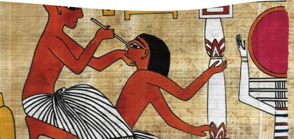
- According to Eber’s papyrus
(1400 BC), the Egyptians
thought that the inner part
of the eye communicated
by a canalicular system with
the inner ear cavities and
the lacrimal cavity - The Egyptians hoped to
restore lost vision by
replacing the inner part of
the eye with a lyophilized
mixture injected into the
inner ear
Ancient Egyptian surgery
- Eber’s papyrus states that the best mixture to replace the
vitreous was also a remedy for total blindness: - “extract of pig’s eye humour 1 part,
- genuine Galenos 1 part,
- yellow ochre 1 part,
- fermented honey 1 part”
- Today, the vitreous is known to provide an optically clear medium,
a mechanical buffer, and a nutrient tissue
Vitreous anatomy
Ophthalmoscopically transparent but structure can be observed with slit lamp
- Rule of “4’s”
- 4 grams
- 4 cc
- Occupies 4/5 of the globe
- 4 x as viscous as water at birth
- Consistency similar too the albumin of an egg
Substructure of the vitreous
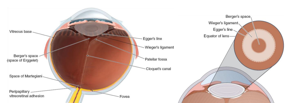
Anatomical openings, spaces an attachments play an important role in the development of pathology.
Vitreous biochemistry
99% water, rest solids
- Acts as a viscous gel or interconnected meshwork
- Gel structure contains long, thick, nonbranching, mostly type 2 collagen fibrils suspended in a network of hyaluronic acid (HA) – type IV collagen – crosslinks
- Proteins (fibrillin & opsin), amino acids, vitamin C (ascorbic acid), and cells (hyalocytes and fibroblasts) are also
found - Worst suggest the adult vitreous body is composed of a number of cisterns
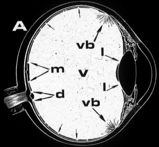
Vitreoretinal interface
- The cortex is composed of two parts;
- anterior cortex/anterior hyaloid face,
begins about 1.5 mm anterior to the ora
serrata - Daicker likened this structure to Velcro –
this explains the strong vitreoretinal
attachment at this site - posterior cortex/hyaloid – 100-110 µm
thick and consists of densely packed
collagen fibrils. - Hogan demonstrated that just posterior to
the ora, heavy bundles of vitreous fibrils
attach to the basal lamina of retinal glial
cells (Müller Cells)
- anterior cortex/anterior hyaloid face,
- There is no cortex over the disk, and the cortex
is thin over the macula and fibers are more
spread out
Vitreoretinal interface – Hyaloid and ILM
- The cortex/hyaloid is firmly attached to the ILM of the retina at the vitreous base, around the disc (Weiss ring), at the vessels, and the foveola at a diameter of 500 µm
- The ILM is 1 – 3 µm thick, consisting mainly of type IV collagen and proteoglycans.
- The ILM can be considered the basal lamina of the Müller cells