

Dr Dirk J. Booysen
Optometrist
Global prevalence of glaucoma and projections of glaucoma burden through 2040
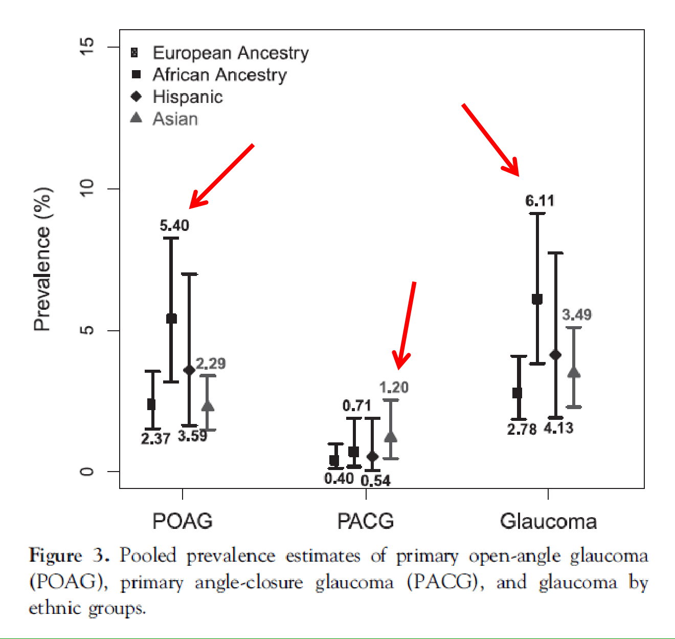
Table 2. Pooled Prevalence (%) and Number of People (Aged 40–80 Years, in Millions) with Primary Open-Angle Glaucoma, Primary Angle-Closure Glaucoma, and Glaucoma in 2013
| World Region | POAG | PACG | Glaucoma (POAG and PACG Combined) | |||
|---|---|---|---|---|---|---|
| Prevalence | Number | Prevalence | Number | Prevalence | Number | |
| Asia | 2.31 (1.44–3.44) | 23.54 (18.32–29.73) | 1.09 (0.43–2.32) | 15.47 (6.26–32.41) | 3.40 (2.26–5.02) | 39.00 (27.78–55.80) |
| Africa | 4.20 (2.08–7.35) | 7.03 (4.25–10.60) | 0.60 (0.16–1.48) | 1.26 (0.34–3.30) | 4.79 (2.63–8.03) | 8.29 (5.16–12.30) |
| Europe | 2.51 (1.54–3.89) | 5.36 (3.99–7.11) | 0.42 (0.13–0.98) | 1.41 (0.43–3.37) | 2.93 (1.85–4.40) | 6.77 (4.94–9.24) |
| North America | 3.29 (1.83–5.53) | 2.97 (1.96–4.29) | 0.26 (0.03–0.96) | 0.39 (0.04–1.38) | 3.55 (1.98–5.81) | 3.36 (2.21–4.94) |
| Latin America and the Caribbean | 3.65 (1.90–6.54) | 5.01 (2.70–8.88) | 0.85 (0.14–3.00) | 1.59 (0.31–5.24) | 4.51 (2.44–7.90) | 6.59 (3.61–11.95) |
| Oceania | 2.63 (1.16–4.83) | 0.20 (0.10–0.33) | 0.35 (0.05–1.15) | 0.05 (0.01–0.16) | 2.97 (1.38–5.23) | 0.25 (0.13–0.42) |
| Worldwide | 3.05 (1.69–5.27) | 44.11 (31.32–60.94) | 0.50 (0.11–1.36) | 20.17 (7.39–45.86) | 3.54 (2.09–5.82) | 64.26 (43.83–94.65) |
PACG = primary angle-closure glaucoma; POAG = primary open-angle glaucoma.
Number of people (aged 40–80 years) in 2013 was estimated on the basis of World Population Prospects: The 2012 Revision from Department of Economic and Social Affairs, United Nations. Worldwide population number (aged 40–80 years) in 2013 was 2.33 billion.
Tham, Yi-Chung et al. Global prevalence of glaucoma and projections of glaucoma burden through 2040: a systemic review and meta analysis, Ophthalmology 2014; 1-10
Projected number of Glaucoma cases
- POAG more prevalent in Africa
- PACG more prevalent in Asia
- Male gender, African ancestry, and urban living were associated with increased risk for POAG development
- Number of patients with glaucoma world wide (40 – 80 year old age group)
- 2013 = 64.3 million
- 2020 = 76 million
- 2040 = 111.8 million (double 2013)
- We need more streamlined screening, treatment and public health strategies
Tham, Yi-Chung et al. Global prevalence of glaucoma and projections of glaucoma burden through 2040: a systemic review and meta analysis, Ophthalmology 2014; 1-10
Key Points
- Severe VF loss leads to profound loss of quality of life
- Moderate VF loss can have implications on daily life
- Early VF loss has little impact on daily life but….
- Early VF loss is the real danger because patients don’t notice their sight loss, so
- Difficult to detect
- Causes them not to adhere to treatment
- Can lead to a catastrophic event (e.g. fall or driving accident)
- We must prevent patients progressing to the other states with treatment
Hyman et al. Ophthalmology 2005;112:1505–13.
Ramulu et al. Curr Opin Ophthalmol 2009;20:92–8.
Lead time gained by OCT in detecting Glaucoma
- In 35% of glaucoma suspects (individuals not yet exhibiting repeatable SAP defects), changes in RNFL with OCT were detectable up to 4 years before visual field defects were detected on SAP (specificity 95% and sensitivity 53%)
- 19% of eyes had abnormal RNFL with OCT up to 8 years prior to visual field defects on SAP(specificity 95% and sensitivity 53%)
(Kuang, Zhang et al. 2015)
RNFL thickness and reaction times
- The authors examined the relationship between RNFL thickness and SAP in patients with glaucoma (as well as a control group) and the reaction time to peripherally displayed targets (of three contrast levels) while performing a simulated driving task.
- RNFL thickness, SAP mean deviation, and age were significantly predictive of reaction times, with worse reaction times observed with low-contrast targets.
- This information suggests that structural measures, including RNFL thickness, can supplement visual field measures to predict which patients may have difficulty with divided-attention tasks, such as driving.
AJ Tatham, ER Boer, PN Rosen, M Della Penna, D Meira-Freitas, RN Weinreb, LM Zangwill, FA Medeiros; Glaucomatous Retinal Nerve Fiber Layer Thickness Loss Is Associated With Slower Reaction Times Under a Divided Attention Task; Am J Ophthalmol 2014 Jul 25;[EPub Ahead of Print],
When Do Patients Realise?
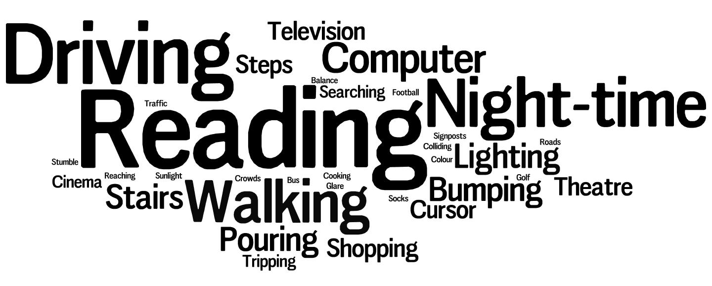
- 50 patients described the situation when they noticed their vision loss
What Do Patients See?
Conclusion
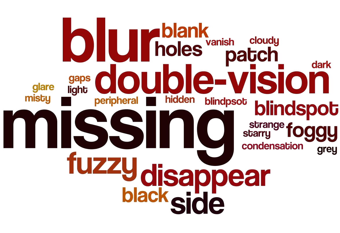
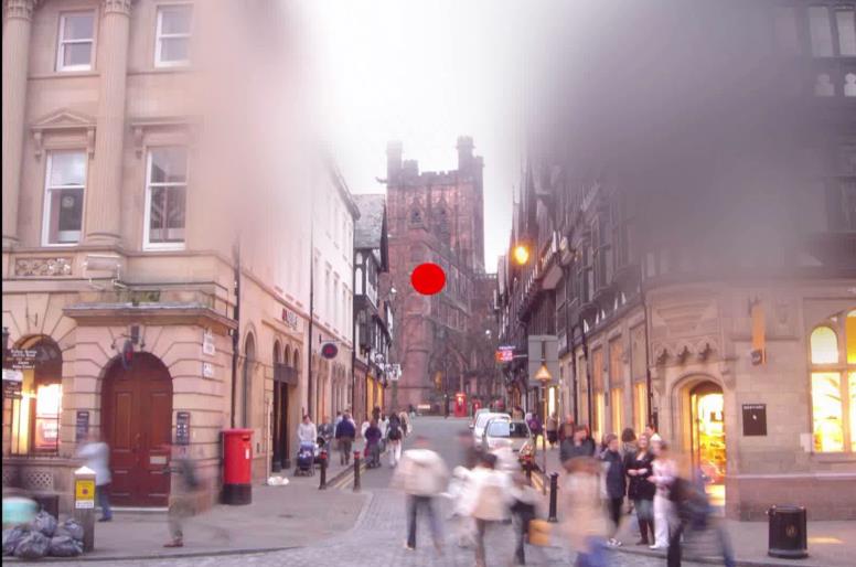
- Patients with glaucoma do not see black areas and most do not have ‘tunnel’ vision
- Important in the context of raising awareness for glaucoma detection and patient education
Treatment strategies
Introduction
- Management of glaucoma should be individualised for each patient
- Clinical evaluation leads to
- Classification
- Risk factor assessment
- Treatment options or observation should be discussed with the patient
- A plan for further visits is recommended
Glaucoma or Not Glaucoma?
- Give your opinion
- Glaucoma or not?
- Treatment?
- Do I need more information?
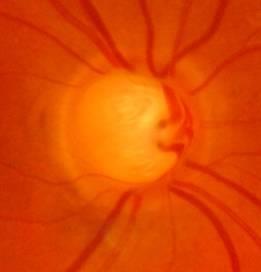
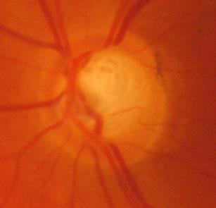
Glaucoma Patients in Everyday Practice
- New patient ‘initial assessment’
- Clinical evaluation
- Risk factors
- Classification
- Stage of the disease
- Treatment decision
- Follow-up
- Patient under follow-up
- Clinical change
- Treatment adjustment
- Follow-up
New Patient: Evaluation
- Patient characteristics
- Age
- Systemic diseases
- Concomitant medications
- Ocular characteristics
- Clinical presentation
- Concomitant eye diseases
- Patient’s expectations and fears
- Mistaken beliefs
- Blindness
- Clinical evaluation
- Visual acuity and refraction error
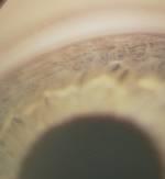
- Anterior segment evaluation
- Intraocular pressure (Goldmann)
- Gonioscopy
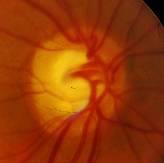
- Posterior pole evaluation (optic nerve head and retinal nerve fibre layer)
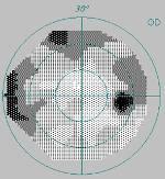
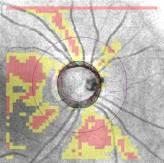
- Visual field and structural assessment
Other useful information
- Known maximum intraocular pressure
- Pachymetry
- Corneal biomechanics
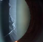
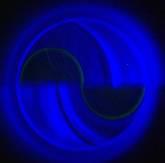
New Patient: Risk Factors
- Assess risk factors for glaucoma diagnosis
- Intraocular pressure
- Age
- Family history
- Race
- Myopia
Cook et al. Can J Ophthalmol 2012;47:223–6.
Uncertain Risk Factors For Glaucoma
In glaucoma, the risk factors below are uncertain and controversial
| Parameter | Trend |
|---|---|
| Vasospastic disease (e.g. migraine, Raynaud’s disease) | Increased incidence and prevalence in NTG |
| Nocturnal (blood pressure) dipping | Increased RoP |
| High blood pressure | Correlates with IOP but not incidence/RoP |
| Ocular perfusion pressure | Uncertain |
| Sleep apnoea | Controversial |
| Diabetes mellitus | Historic |
New Patient: Decision-Making
- Should you treat?
- Yes as a rule, if
- Established glaucoma
- Ocular hypertension with confirmed high IOP
in which optic nerve damage is likely to occur
- Consider not treating if
- The diagnosis is not clear (‘equivocal disk’)
- Ocular hypertension suggests low risk
- Glaucoma suspects
- Mild glaucoma in a very old patient should be discussed with the patient and family
- Low tension glaucoma
- Primary angle-closure suspects
- It is important to set treatment objectives
- Efficacy (target intraocular pressure)
- Safety
- Education of the patient
EGS guidelines, 3rd edn, 2008
Tatham et al. Eye 2013;27:1293–8.
First-Choice Treatment
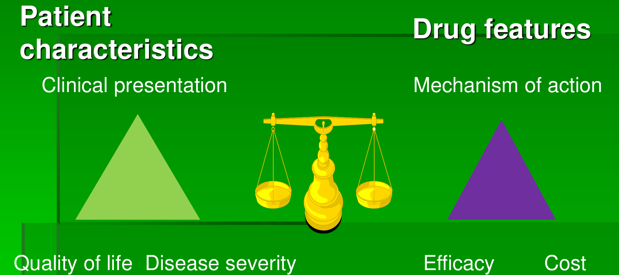
Treatment Options: First-line/First-choice Drugs
| Drug | IOP lowering | Main limitations | ||
|---|---|---|---|---|
| Peak | Trough | Local | Systemic | |
| Prostaglandin analogues | 33% | 28% | Conjunctival hyperaemia, pigmentation, eyelash growth | Dyspnoea (shortness of breath) |
| Beta-receptor antagonists | 27% | 26% | Conjunctival hyperaemia | Asthma, arrhythmia, libido |
| CAI A-2 | 20% | 17% | Burning, stinging, bitter taste | Kidney or liver damage |
| Selective adrenergic agonists | 25% | 18% | Ocular hyperaemia, lid retraction, mydriasis | Fatigue, drowsiness, hypo-/hypertension |
Van der Valk et al. Ophthalmology 2005;112:1177–85
Avoid Decisions Based on…
- A single intraocular pressure measure
- A visual field change without confirmation
- A structural change without confirmation with the same machine
- Be aware of structural imaging artefacts!
- Avoid making decisions without performing visual field tests
Conclusions
- Consider the patient as a whole
- Take the time to document clinical findings
- Take the time to document progression, if any
- Evaluate the benefit versus safety of medication when considering therapy
- Regularly re-evaluate your strategy
To Treat or Not to Treat?
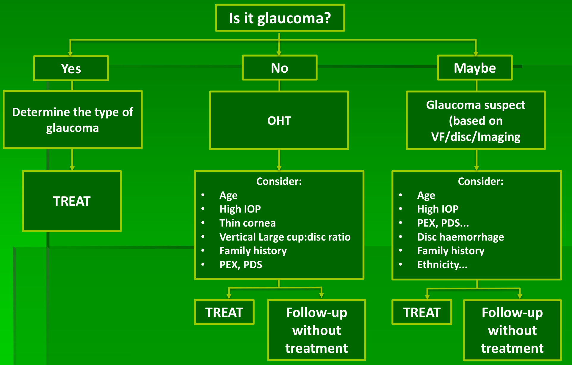
Step 1. Before Initiating Medical Treatment Check Systemic and Ocular Safety
Systemic safety
| Asthma, bradycardia | Nephrolithiasis | Children | Pregnancy |
|---|---|---|---|
| Avoid • Beta-blockers • Fixed combinations incl. beta-blockers | Caution • Carbonic anhydrase inhibitors* | Avoid • Alpha-agonists | Caution • All drug classes* |
Ocular safety
| Ocular surface disease | Corneal endothelial disease | Risk of cystoid macular oedema |
|---|---|---|
| Caution • BB/ Preserved drops | Caution • Topical carbonic anhydrase inhibitors | Caution • Prostaglandin analogues |
* Consider risk-benefit balance carefully
*Carlsen J(1), Durcan J, Zabriskie N, Swartz M, Crandall A Arch Ophthalmol. 1999 Aug;117(8):1087-8.
Step 2. First-Choice Medical Treatment According to Baseline IOP, Level of Damage and Drug Efficacy Profile
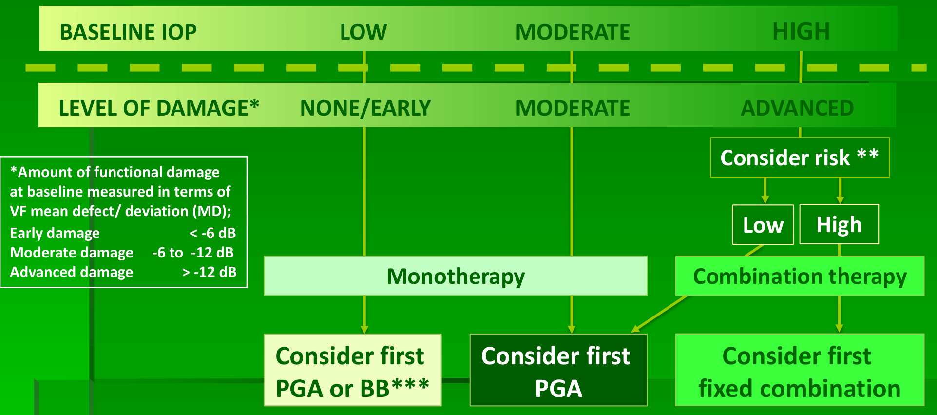
** Very high IOP; (strong evidence) age, level of damage, exfoliation; (weak evidence)
As a rule, lower pressures recommended in: younger patients, advanced damage, presence of exfoliation and thinner cornea
***Avoid BB in pts with low BP or slow heart rate.
Adopted after EGS guidelines 2008
How to Treat? Specific Conditions
Individual Characteristics / Pathogenesis / IOP-lowering demands
Consider prioritising the following options in specific conditions:
| Pseudoexfoliation | Angle-closure | Patients at high risk for progression* Greater IOP reduction (%) required |
|---|---|---|
| Mild: Laser trabeculoplasty/ medical treatment Advanced: low threshold for intensive treatment, in addition careful monitoring | 1° PI 2° Consider cataract surgery (or combined surgery depending on IOP, level of damage & synechia) | Combination therapy Surgery |
*High risk patients: advanced disease, very high IOP, in combination with other risk factors, e.g. family history
Adopted after EGS guidelines 2008
Step 3. Responses and Further Actions After Starting First-Choice Medical Therapy
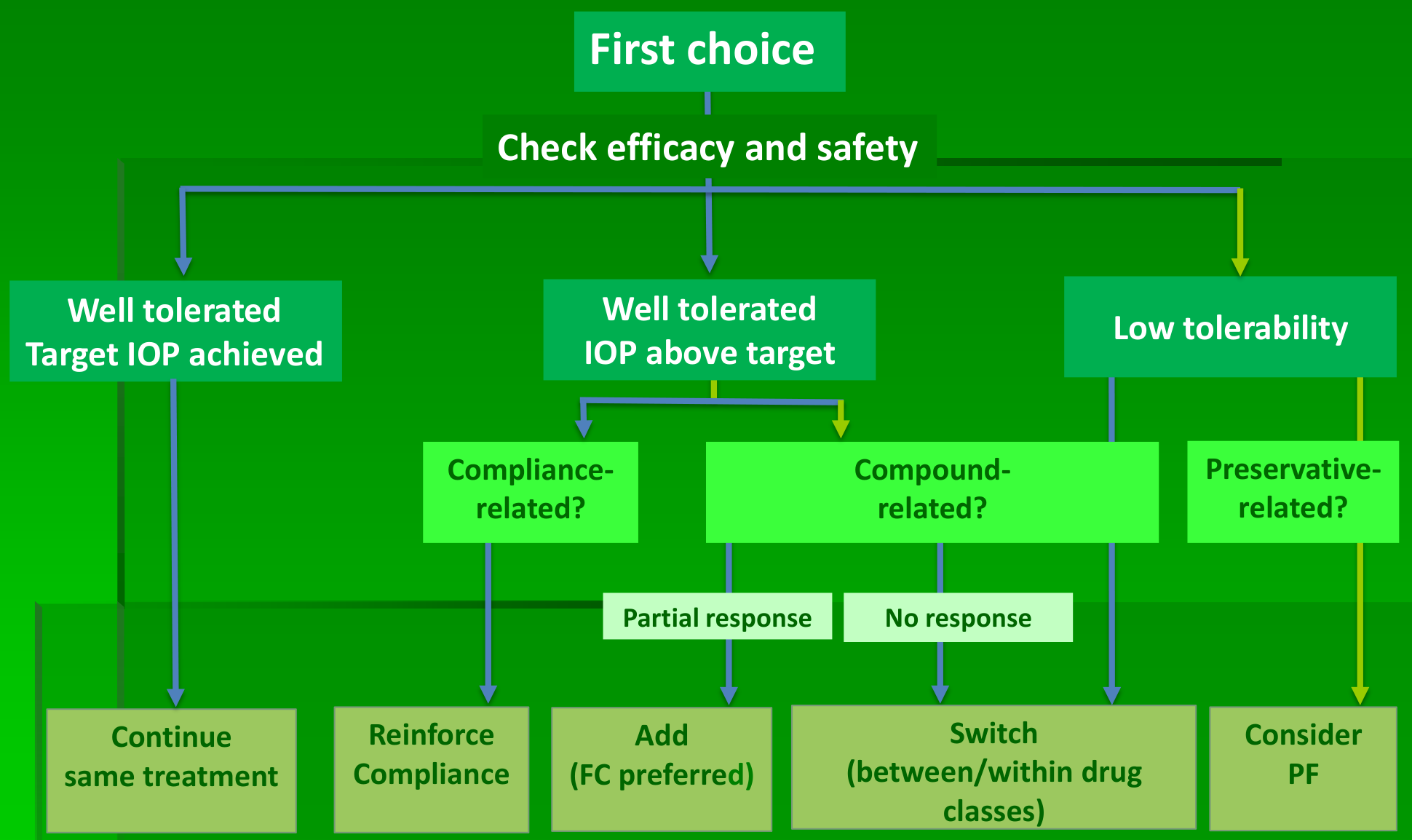
Assessment of Compliance/ Understanding/Tolerability: ‘Tell Me…’
Tell me please…
- How do you feel about your drops?
- How and when do you put your drops in?
- What time did you last put in your eyedrops?
- How do you remember when to use them?
- What are the main difficulties you experience with your drops?
Talking about compliance is always important. Use open questions.
Target IOP Not Reached: Considerations
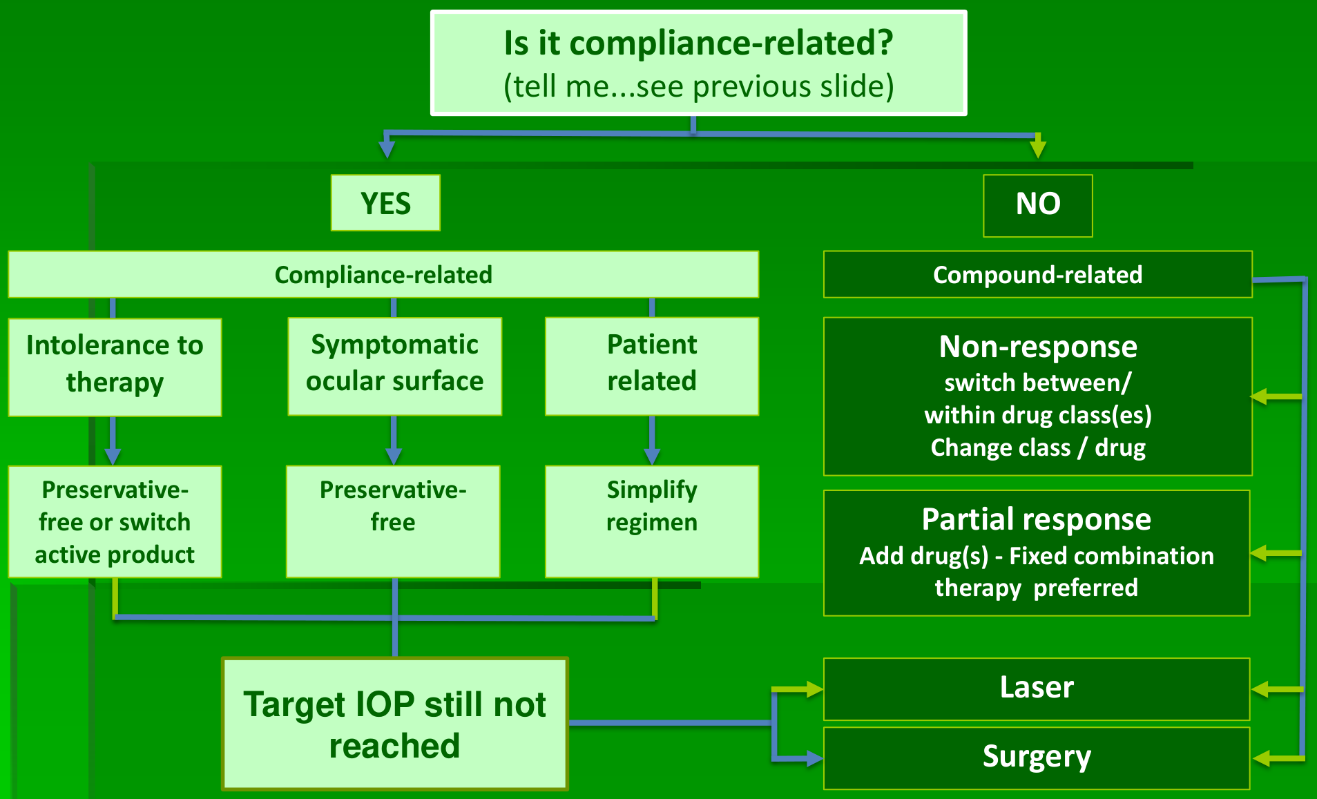
In patients who are stable despite higher-than-target IOP: consider accepting higher target!
The influence of each mmHg IOP reduction on functional progression
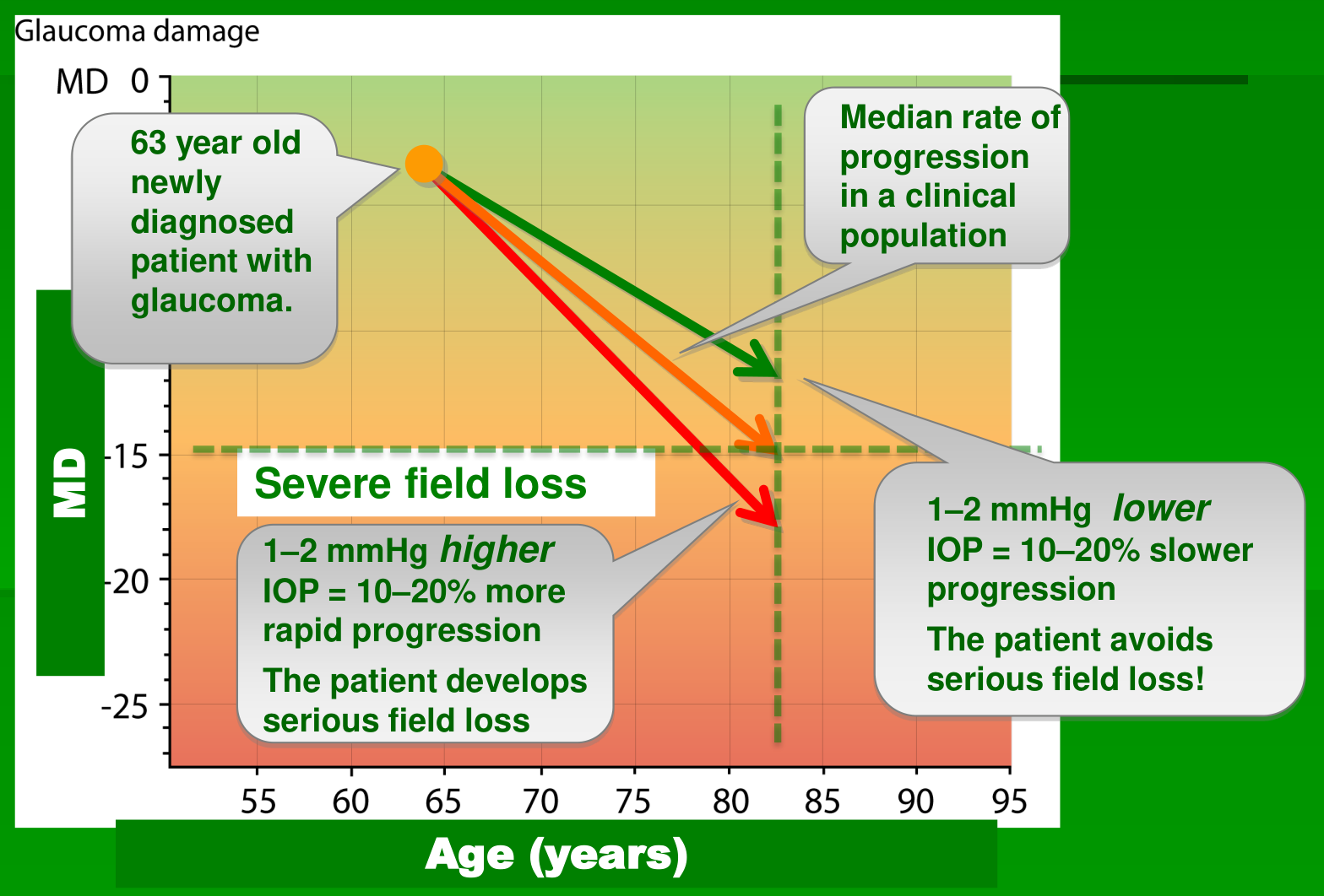
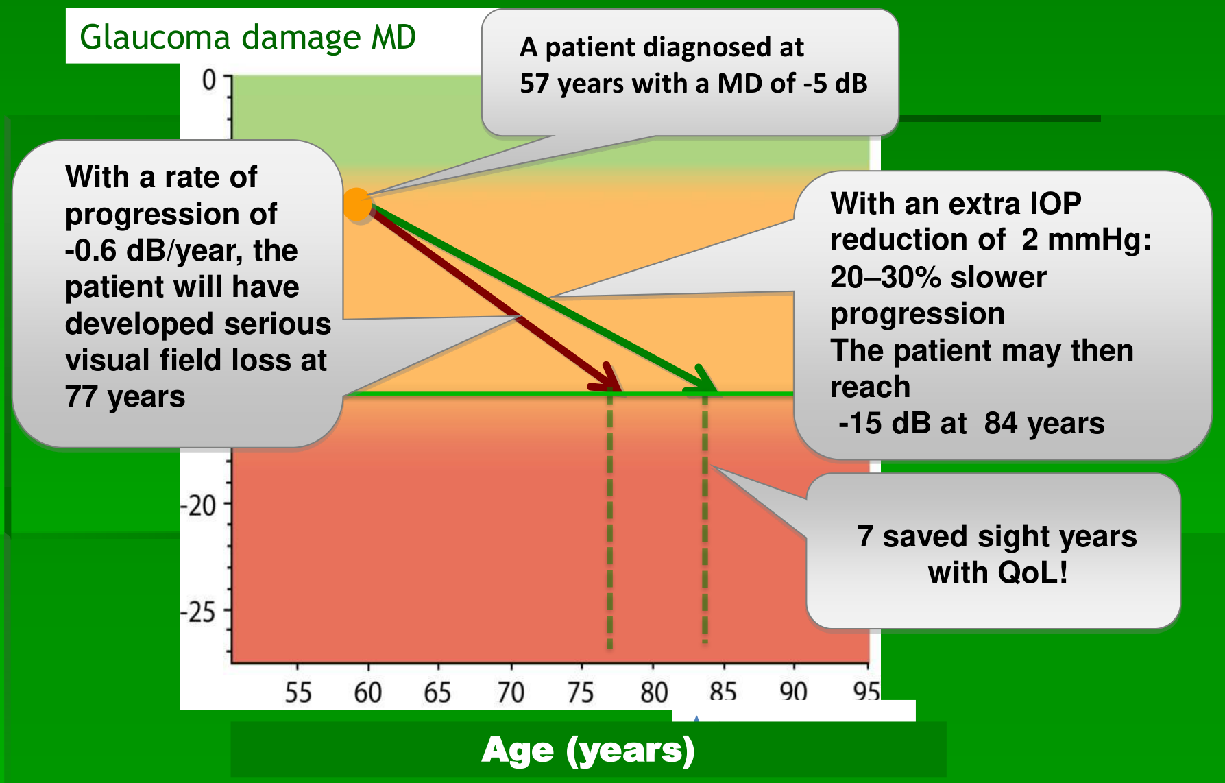
By Changing the Visual Field Rate of progression, a patient’s QoL can be preserved for longer
Treatment, Key Points
- Look at each patient as an individual
- Look for individual risk factors
- Determine the stage of the glaucoma
- Evaluate the rate of progression
- Identify fast progressors
- Adjust target intraocular pressure – sight saving years?
- Inform and try to educate the patient and family
Thank you