
Dip Optom (SA), TMOD (US), CAS (US), FC Optom (UK), PhD (Aston), FSLS, FOA(SA), FICLS
Member of Aeromedical Committee SACAA
dirk@dirkbooysen.co.za
All DAMES should understand the visual environment in which aviation personnel work!
The Visual System in the Flight Environment
Environmental factors influencing visual function
- Altitude and Hypoxia
- Reversed light distribution, high altitude sky is black not blue
- Low humidity, contact lens and dry eye problems
- Empty field myopia up to 1.5 diopters
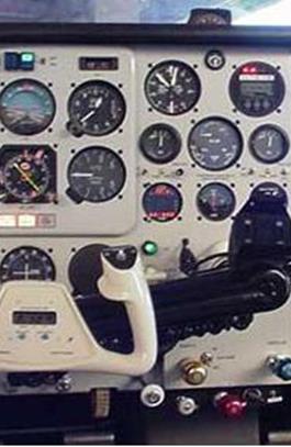
- Cockpit illumination
- Low level illumination reduce DOF: Dilated pupils, increased presbyopia, reduced visual acuity, and colour vision
- Red light cockpit illumination causes problems with colour vision and may increase hyperopia
- Speed
- Dynamic visual acuity and threshold for angular motion more important than static visual acuity – affects reaction time
- Relative decrease in dynamic visual ability after the age of 50yrs is of concern in older pilots
- Acceleration
- Effects of G-Force, red out, grey out and black out
- Vibration
- Especially within the 22 – 64 Hz range may cause difficulty in reading instruments or printed material (mostly in helicopters)
- Vibration in the range of 2–10 Hz encountered in turbulence and rough runways has a significant detrimental effect on visual performance
- Ergonomics and cabin environment
Human factors such as stress and fatigue
Visual Flight deck & ATC tasks
Distance visual tasks
Intermediate and near visual tasks
Spatial orientation
Processing colored information
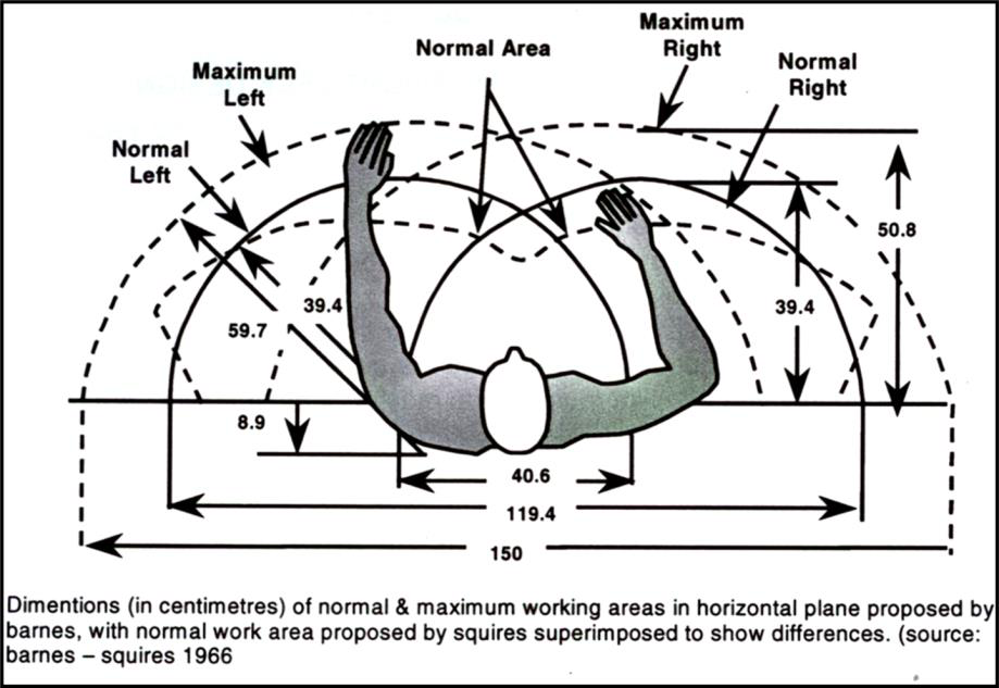
Ave working distance 50cm
ATC specific visual tasks
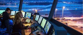
- ATC’s deal with, distance, intermediate, and near tasks
- Working in the tower requires good distance visual acuity to identify aircraft in the circuit
- Often the radar images use high resolution color coding to identify different aircraft
What is Required in Aviation Vision Examination?
- Overall ocular health – history including any surgery
- Examination of the anterior segment – pay attention to the cornea, look for scars that may indicate prior surgery
- Examination of the posterior pole – ophthalmoscopy
- Visual acuity, monocular and binocular, unaided and aided at distance, near and intermediate testing distances
- Refractive error – spectacle correction and or contact lens correction
- Binocular vision and muscle coordination
- Peripheral vision and visual fields
- Colour vision
Medical certification, 4 classes
| Class 1 | Class 2 | Class 3 | Class 4* |
|---|---|---|---|
| ALTP | Private pilots | ATC | Recreational pilots |
| CPL | Student pilots | Drone Pilots | ATC assistants |
| Flight test ratings | Cabin Crew Members | *Medical examination can be performed by any medical doctor | |
| Flight engineers |
General – Visual System
An applicant may not have –
- Any condition or congenital abnormality of either eye or its attachments likely to impede the safe exercise of the privileges of the licence;
- Any abnormality of visual fields or significant defect of binocular function;
- Any manifest squint, or large errors of eye muscle balance (phoria);
- Any anatomical or functional monocularity or substandard vision in one eye at initial issue of a Class 1 medical certificate. However, medical conclusion may permit experienced licence holders who develop monocularity or substandard vision to be granted a medical certificate with appropriate restrictions after an adaptation period of at least six months following the loss of vision.
Visual Standards
| Class 1 | Class 2 | Class 3 | Class 4 | |
|---|---|---|---|---|
| Distance vision | At 6 meters, 6/9, 20/30, 0.66 in each eye or better, and binocularly 6/6, 20/20, 1.0 or better with or without corrective lenses | At 6 meters, 6/12, 20/40, 0.50 in each eye or better, and binocularly 6/9, 30/30. 0.66 or better with or without corrective lenses | At 6 meters, 6/12, 20/40, 0.50 in each eye or better, and binocularly 6/9, 20/30, 0.66 or better with or without corrective lenses | At 6 meters 6/12, 20/40 , 0.50 in each eye or better, and binocularly 6/12, 20/40, 0.50 with or without corrective lenses |
| Intermediate vision | Read N14, 6/30, 20/100, 0.20 at 100 cm or have equivalent monocular acuity of 6/24, 20/80, 0.25 at 100cm | Read N14, 6/30, 20/100, 0.20 at 100 cm or have equivalent monocular of acuity 6/24, 20/80, 0.25 at 100cm | Read N14, 6/30, 20/100, 0.20 at 100 cm or have equivalent monocular acuity of 6/24, 20/80, 0.25 at 100cm | Read N14, 6/30, 20/100, 0.20 at 100cm or have equivalent monocular acuity of 6/24, 20/80, 0.25 at 100cm |
| Near vision | Read N5, 6/9, 20/30, 0.66 at a distance of 33 cm or have equivalent monocular acuity of N6, 6/12, 20/40, 0.50 at 33cm | Read N5, 6/9, 20/30, 0.66 at a distance of 33 cm or have equivalent monocular acuity of N6, 6/12, 20/40, 0.50 at 33cm | Read N5, 6/9, 20/30, 0.66 at a distance of 33 cm or have equivalent monocular acuity of N6, 6/12, 20/40, 0.50 at 33cm | Read N5, 6/9, 20/30, 0.66 at a distance of 33 cm or have equivalent monocular acuity of N6, 6/12, 20/40, 0.50 at 33cm |
If uncorrected distance VA is worse than 6/60 in either eye, the applicant must provide a full ophthalmic report prior to initial assessment, and then every 5 years thereafter
Diopter Limits
- A need for corrective lenses for either eye within the range of plus or minus 5 dioptres (spherical equivalent) may be accepted, provided that the visual acuity without correction is not worse than 6/60 in each eye separately.
- Spectacle lenses outside this range are not routinely acceptable, but medical conclusion may permit an applicant to be assessed as fit on production of satisfactory specialist reports.
- The medical certificate will be, where appropriate, endorsed with the following:
- “Contact lenses only must be worn”; and
- “Spare spectacles must be readily available”.
Minimum restrictions for monocularity, amblyopia and substandard vision in one eye
- Any anatomical, functional monocular or substandard vision in one eye at initial issue of medical certificate
- Monocularity means vision can not be corrected to 6/24, 20/80, 0.25
- Substandard vision in one eye means central vision better than 6/24, 20/80, 0.25 but worse than 6/9, 20/30, 0.66 with normal visual fields
- Experienced license holders that develop monocular or substandard vision in one eye may be granted certification with appropriate restrictions
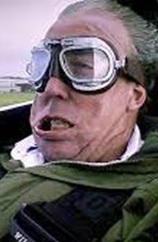
- Protective goggles in open cockpit aircraft must be used
- Accompanying pilots must be made aware of the holders monocular or substandard vision in one eye
- Not valid for PIC by day or night until an appropriate flight test has been completed
- No active ocular pathology present
- Distance vision (corrected or uncorrected) must be 6/6, 20/20, 1.0 or better and near vision 6/9, 20/30, 0.66 or better
Contact Lenses
Applicants may use contact lenses to meet visual requirements, provided that:
- the lenses are monofocal/single vision and non-tinted (no bifocal or monovision allowed);
- the lenses are well tolerated;
- And a pair of suitable correcting spectacles is kept readily available during the exercise of the licence privileges.
- Applicants who use contact lenses may not need to have their uncorrected visual acuity measured at each re-examination provided the history of their contact lens prescription is known.
- Applicants with a large refractive error shall use contact lenses or high-index spectacle lenses to correct their vision to acceptable standards
- Finally, the applicant shall be required to have normal fields of vision as well as normal binocular function.
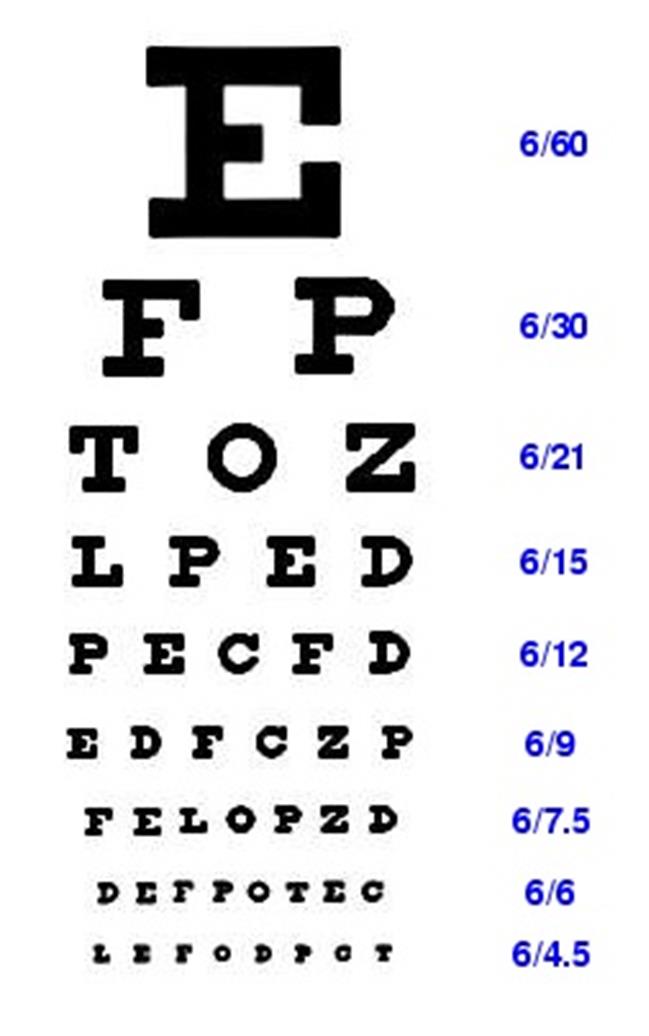
| Snellen Row | Size of Letters |
|---|---|
| 6/60 | 88.7 mm |
| 6/40 | 59.1 mm |
| 6/30 | 44.4 mm |
| 6/24 | 35.5 mm |
| 6/18 | 26.6 mm |
| 6/15 | 22.2 mm |
| 6/12 | 17.7 mm |
| 6/9 | 13.3 mm |
| 6/6 | 8.9 mm |
| 6/4 | 5.9 mm |
VA measures taken at distances beyond 2 meters are directly comparable and should show consistent values independent of the distance. Diseases and refractive errors were not shown to impact on the VA results at different distances
November 2018. Effects of Testing Distance on Visual Acuity Measures. [online]
Distance Visual Acuity Testing
- Standard Snellen Chart with either letters or Landolt C symbols
- Preferred – 6meter distance but specialized charts for 3meter test distances acceptable
- Adequate illumination – 100 Watt at 1.2meters onto chart (1600 Lumens), shielded from applicant
- Room illumination should be dark
- Unaided, aided vision taken monocularly as well as binocularly
- Can use either spectacles or contact lenses
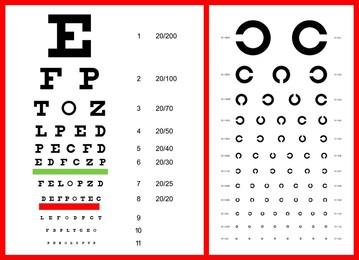
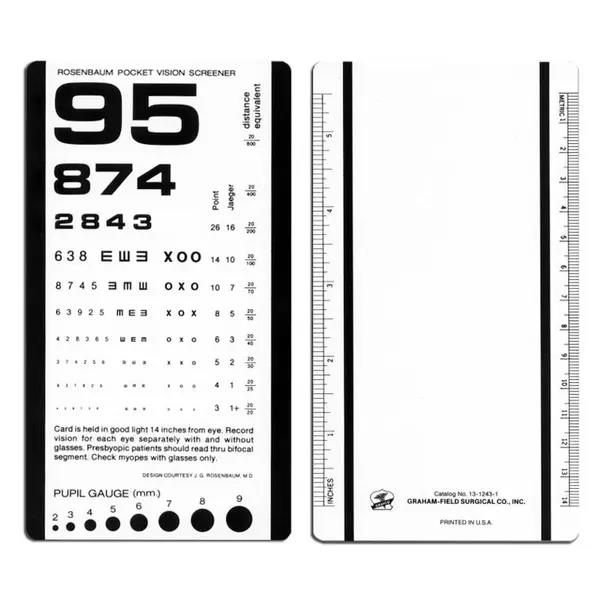
Rosenbaum Pocket Vision Screener
- The Pocket Vision Screener was found to have 81% sensitivity, 94% specificity. The positive and negative predictive values were found to be 91% and 87%, respectively.
- Therefore, this can be used as quick and accurate tool to screen subjects with visual acuity worse than 6/9.
- The Pocket Vision Screener is a compact and simple tool to use in addition to being very cost effective.
Raja M, Ramamurthy D, Srinivasan K, Varadharajan LS. Development of Pocket Vision Screener and its effectiveness at screening visual acuity deficits. Indian J Ophthalmol. 2014 Dec;62(12):1152-5. doi: 10.4103/0301-4738.149137. PMID: 25579360; PMCID: PMC4313496.
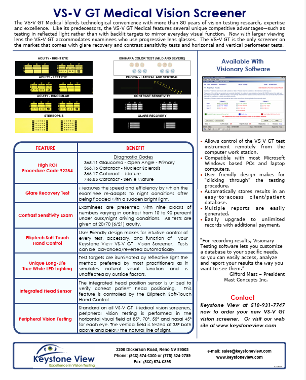
Keystone Vision Screener (Orthorator)
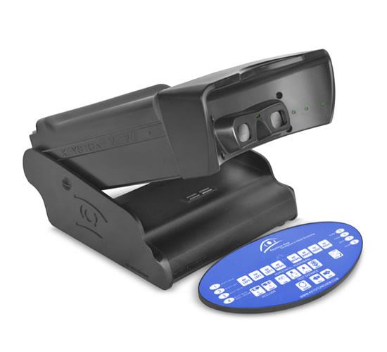
$3600.00?
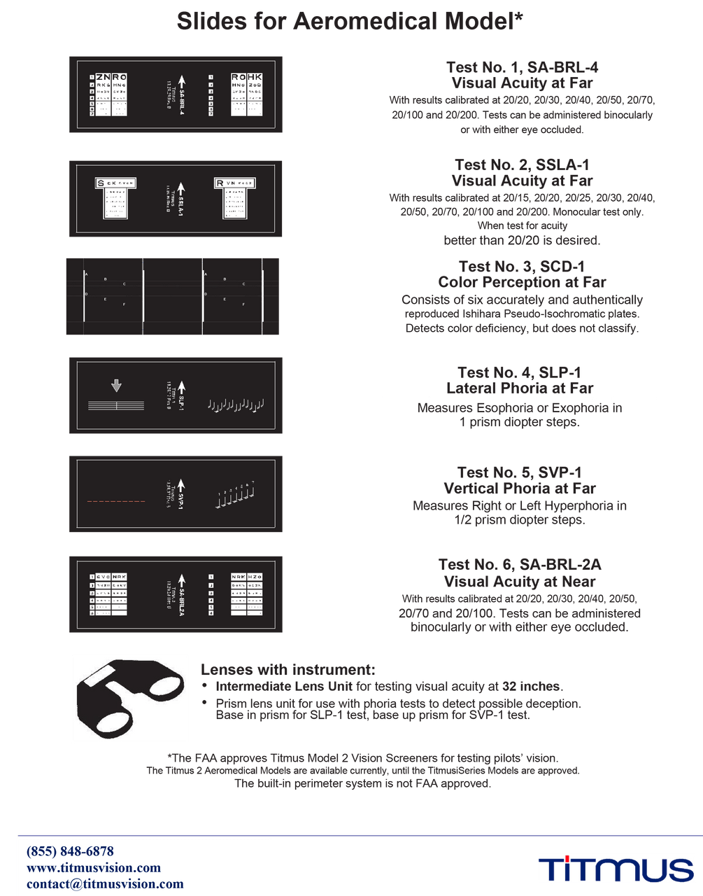
Titmus V2 Vision Screening with Aeromedical Test Slide Package
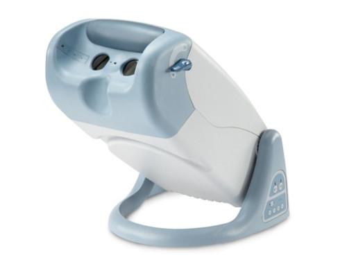
$3625.00
Problems with Keystone and Titmus Stereoscope Screening Tests
- High False Positive Rate: They identify individuals as having a vision problem when they do not.
- Calibration and Training: For the tests to be valid and accurate, the instruments must be properly calibrated and the screening personnel must be carefully trained on the correct procedures and instructions provided by the manufacturer
- Limited Scope: Vision screening is not a diagnostic tool; it identifies potential issues that require further examination.
- High Costs: Compared to simpler methods like the Snellen chart and Pocket screener, these stereoscopes have higher equipment and maintenance costs, contributing to the overall cost of vision screening programs.
- Inappropriateness for Certain Age Groups: Some protocols suggest they are not suitable for children five years and younger.
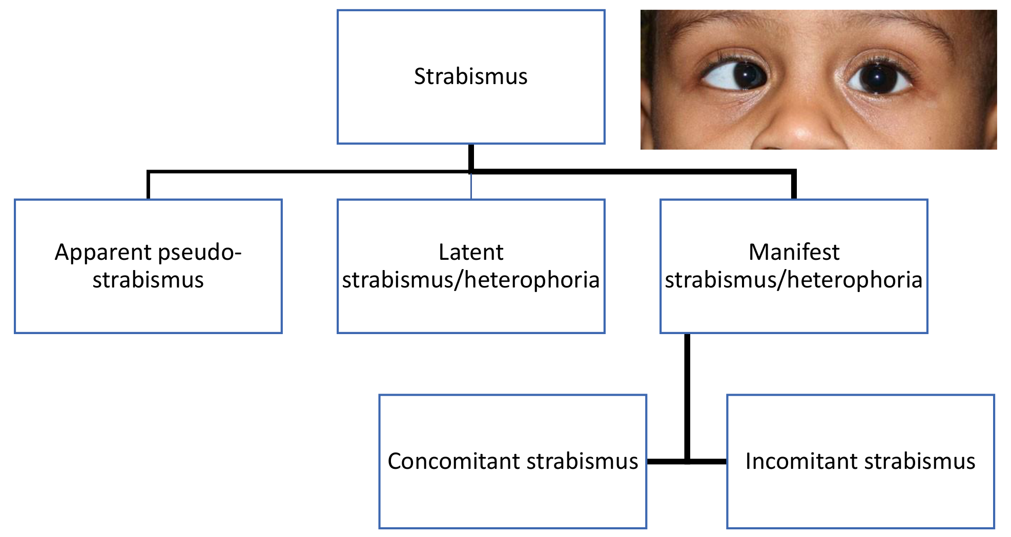
Latent Strabismus/Heterophoria
- Heterophoria is a condition in which the eyes deviation is kept latent by fusion – in other words the deviation is compensated
- When fusion breaks down, one eye deviates and the strabismus becomes manifest – in other words the deviation is decompensated
Heterophoria Types, Features and Causes
| Type | Details | Causes | |
|---|---|---|---|
| Esophoria Eye moves nasally under cover | Convergence excess | Greater at near than at distance | • Anatomical (narrow PD) • Refractive (high hypermetropia) • High AC/A ratio • Weak divergence (negative fusional reserve) |
| Divergence weakness | Greater at distance than at near | ||
| Non-specific | Similar angle at near and distance | ||
| Exophoria Eye moves temporally under cover | Convergence weakness | Greater at near, often but not always with convergence insufficiency | • Anatomical (wide PD) • Refractive (myopia, presbyopia) • Weak convergence (positive) fusional reserves • Age |
| Divergence excess | Greater at distance than near | ||
| Non-specific | Similar angle at near and distance | ||
| Vertical phoria Eye moves up or down under cover | Hyper/hypo | One eye rotates upwards and the other downwards under cover | • Refractive (high myopia) |
| Alternating hyper/hypo | Either eye moves upwards/ downwards under cover | • Weak vertical fusional reserves • Anatomical (abnormal extraocular muscles) • Incomitancy – extraocular muscle anomaly | |
| Cyclophoria Either eye “wheel rotates” under cover | Incyclophoria | Upper end of vertical axis is nasal | • Oblique astigmatism |
| Excyclophoria | Upper end of vertical axis is temporal | • Incomitancy |
SACAA Minimum standards for heterophoria
- SACAA – Acceptable limits are DV – 5∆ exophoria, 10∆ esophoria, 2∆ hyperphoria, 1∆cyclophoria, measured with best spectacle correction
- Ernstig’s Aviation Manual – DV 6∆ esophoria, 8∆ exophoria, 1∆ hyperphoria and NV – 6∆ esophoria, 16∆ exophoria, 1∆ hyperphoria
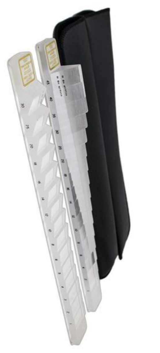
Heterophoria Evaluation – Maddox Rod
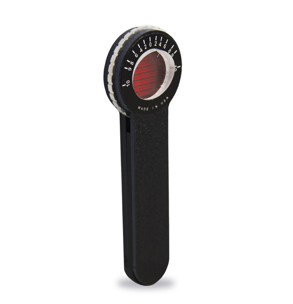
Less than $10.00
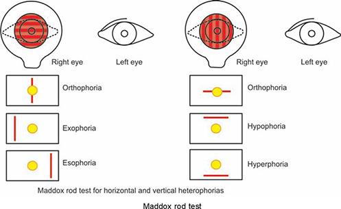
Cover Test and Alternating Cover Test
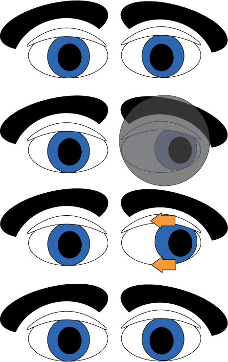
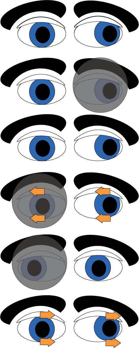
Symptoms of Decompensated Heterophoria
- Headaches: horizontal phoria – frontal headache and vertical – occipital headache
- Asthenopia – group of symptoms including eye ache, fatigue, general soreness and discomfort
- Blurring of print and difficulty changing focus from near to far
- Intermittent diplopia after intense near work
- Intermittent confusion or distorted vision – letters and words appear to move, loose place while reading
- Adaptation by abnormal head posture, closing one eye while reading
Causes of Heterophoria Decompensation
| Optical | Medical | Environmental |
|---|---|---|
| Uncorrected refractive error • Esophoria decompensated by uncorrected hypertropia • Exophoria decompensated by uncorrected myopia | Poor health, fatigue, worry and anxiety • Reduces fusional reserves • Reduces amplitudes of accommodation and subsequent accommodative convergence | Increase in ocular activity • Change in occupation • Extended period of work at a particular distance |
| Over/under corrected refractive error • Reduces the acuity and dissociates the eyes • More pronounced where one eye is affected more than the other | Traumatic injuries • Head trauma can result in temporary or permanent reduction in fusion | Poor working environment • Reading at too close a distance for long periods • Poor illumination or contrast of visual task |
| Poorly fitted spectacles • Particularly with high refractive errors that can induce unwanted prismatic effects | Adverse effects of drugs • Certain anti-hypertensive and anti- depressant agents can reduce accommodation • Alcohol has been found to reduce horizontal fusional reserves | Playing games • Video games that involve rapid, repeated pursuit fixation movements • ‘Magic Eye’ 3D auto stereograms that dissociate and disrupt accommodation and convergence • VR or virtual reality devices |
| Anisometropia • Difference in image size between the eyes (aniseikonia) makes fusion difficult. | Short period of occlusion • Patch worn over one eye following a corneal injury • Patch worn in childhood for the treatment of amblyopia | Night driving • Reduced visual information prevents binocular functioning between the eyes |
| Extensive visual field loss • Glaucoma reduces binocular matching between the eyes • Advanced AMD in one eye | Extensive monocular viewing • Jewellers • Watch makers |
Stereopsis/Depth Perception
- Awareness of the relative distances of objects from the observer by means of binocular vision only – based on retinal disparity
- Good test to determine if person is binocular
- Normal 5 -15 seconds of arc, deteriorates with age and is correlated with Snellen acuity
- Stereopsis only effective < 500 meters – some sources say < 100 meters?
- Reduced stereopsis and abnormal convergence not interfering with near vision, as well as ocular misalignment where the fusional reserves are sufficient to prevent asthenopia and diplopia need not be disqualifying!
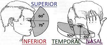
Visual Fields
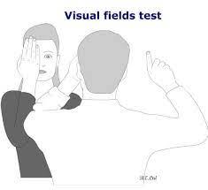
Confrontation Test
- Effective and efficient, and costs nothing but a little time
- The object of this test is to compare the examiners visual field with that of the subject
- Start with the temporal field of the right eye using your left hand. Bring the hand or object slowly in from the temporal field until the subject can see the object or fingers
- Now the nasal field can be tested by using the right hand
- Repeat for the left eye
Color Perception Standards – SACAA
- Applicants must demonstrate ability to readily perceive colors that necessary for the safe performance of duties
- Ishihara 24 (or 38) plate done in daylight or light source (C, D) specified by the ICI
- Considered passed if less than 2 errors in plates 1-15(24) and 4 errors in plates 1-21(38)
- Failing the 24 or 38 plate Ishihara test, assessed fit if they pass CAD – Color Assessment Diagnosis
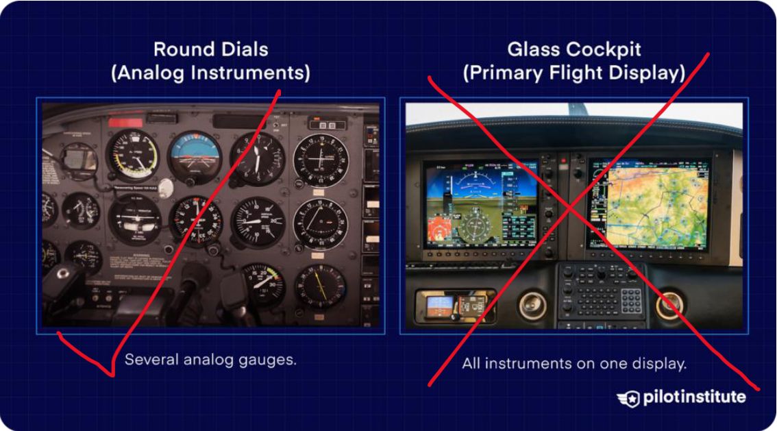
- Class 1, class 2 with CVD will be allowed with restrictions on PPL, no night flight, no glass cockpit
Colour in Context
Visual perception of colour involves a complex series of events and is affected by many variables such as:
- Level of ambient illumination
- Stimulus brightness, saturation
- The influence of adjacent colours, stimulus size, shape, texture and duration
- Visual fatigue and afterimages
- The integrity of the visual system (as affected by age, disease and drug usage)
Young-Helmholtz trichromatic theory of colour vision
- 3 types of retinal cones containing, red sensitive (L-Cone – long wave), green sensitive (M-Cone – medium wave), and blue sensitive pigment (S-Cone – short wave) – trichromatism
- The mean L:M:S cone densities are taken to be in the ratio of about 40:20:1 (Kremers et al., 2000; Walraven and Bouman, 1966)
- Colour vision is limited to stimuli seen within about 40° of the visual axis (Hurvich, 1981); outside this area vision is virtually monochromatic and used mainly for the detection of movement
- Color vision defects occur when there is a deficiency in one or more of the three cone pigments
ADLER, F. H. & HART, W. M. 1992. Adler’s Physiology of the Eye: Clinical Application, Mosby Year Book.
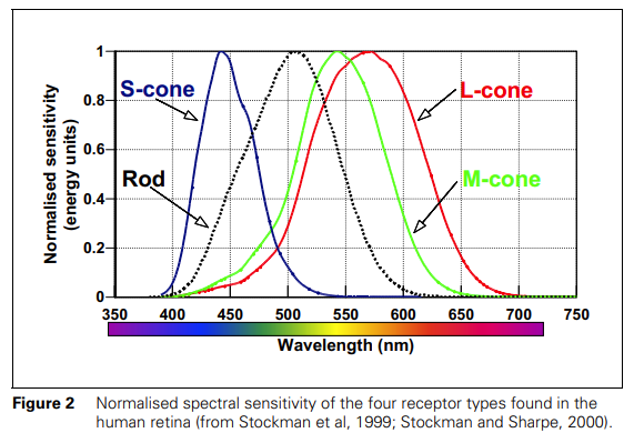
Color Vision Defects
- Most common form is anomalous trichromatism where the individual has all three types of pigment, but one is deficient to some degree
- In dichromatism the individual only has two types of pigment
- Monochromatism – extremely rare and associated with severe visual problems
- Rod monochromats
- Cone monochromats
ADLER, F. H. & HART, W. M. 1992. Adler’s Physiology of the Eye: Clinical Application, Mosby Year Book.
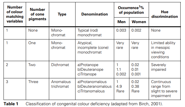
CVD – Dichromatism
- Dichromats
- Protanopia, deficiency in red receptor cones, (severe red / green defect) – blue-green and red seen as grey
- Deuteranopia, deficiency in green receptor cones, (severe red / green defect) – purple-red and green seen as grey
- Tritanopia, Deficiency in blue receptor cones, (severe blue / yellow defect) – confuse blue with green and yellow with violet
All dichromats are at risk for temporary monochromatic vision through tinted glass or spectacle lenses
ADLER, F. H. & HART, W. M. 1992. Adler’s Physiology of the Eye: Clinical Application, Mosby Year Book.
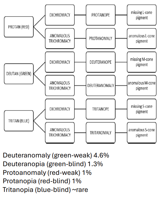
CVD – Trichromatism – most common defect
- Anomalous Trichromats – milder than dichromats
- Protanomaly deficient in red receptor cones, (“blue green and red seen as greyish indistinct color”)
- Deuteranomaly deficient in green receptor cones, (“purple red and green seen as indistinct greyish color”)
- Tritanomaly deficient in blue receptor cones, (“confuse blue with green and yellow with violet, less severe than tritanopia”)
ADLER, F. H. & HART, W. M. 1992. Adler’s Physiology of the Eye: Clinical Application, Mosby Year Book.
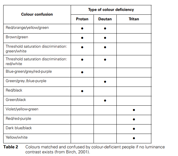
Prevalence of congenital CVD
Green weak is most common colour vision defect in males
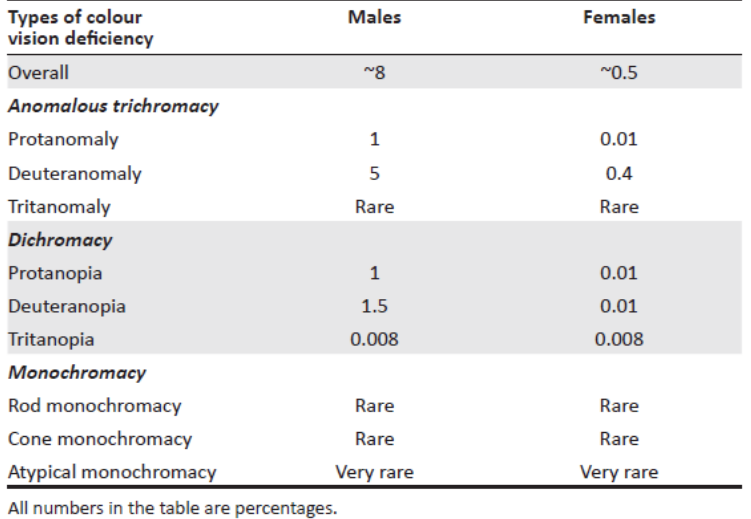
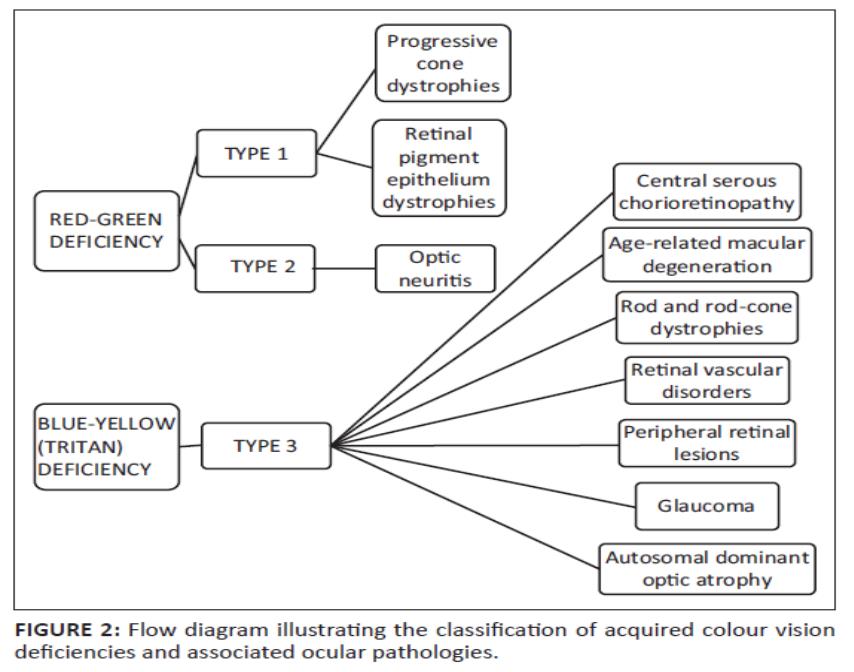
Acquired CVD
Generally;
- Optic nerve related pathology – Red Green Defects
- Retinal and vascular disorders – Blue Green Defects
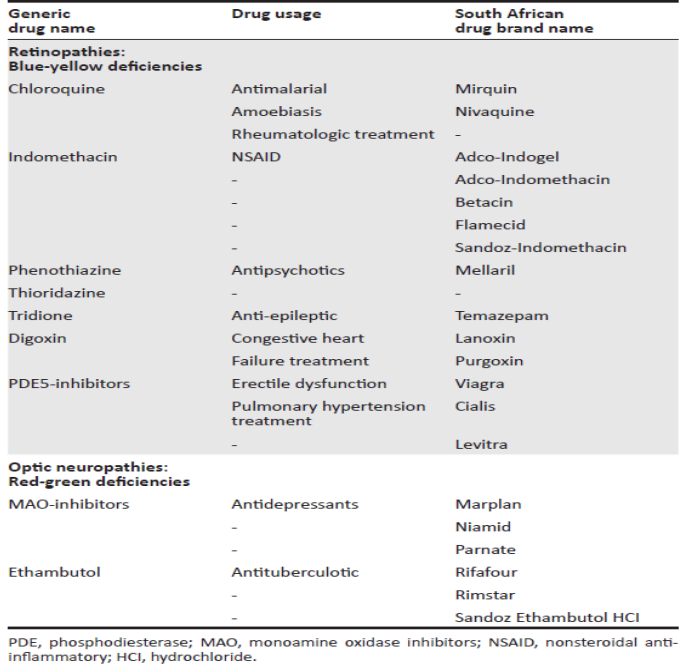
Drug induced CVD
Evaluation of CVD
- CVD can be assessed qualitatively and quantitatively
- Efficiency of the tests are based on their sensitivities and specificities
- No single test has ever reliably matched colour vision deficiency with performance degradation on specific in-flight tasks
- Colour environment in modern cockpits more challenging than before – more appropriate screening tests are required
- Six main categories
- Plate tests – Ishihara is the gold standard for screening
- Arrangement tests
- Lantern tests
- Anomaloscope
- Electroretinography
- Computer based colour vision tests – CAD,
Plate Tests – Ishihara Test
- Design based on the plates of Stilling
- The figure and background are made up of discrete discs that vary in size and luminance to ensure that the figure can be identified only by its chromatic difference from the background and not from a difference in the perceived luminance
- The test is carried out at 66 cm, comprising 24 or 38 plates, and the observer is given four seconds to identify the numerals or pathways
- The plates are divided into transformation, vanishing, hidden and classification plates
- IT should be used only as a pass fail test to detect red-green CVD
- IT has no tritan or YB plates
- Tendency to pass mild deutans and to fail some normal trichromats – (15 – 19%)
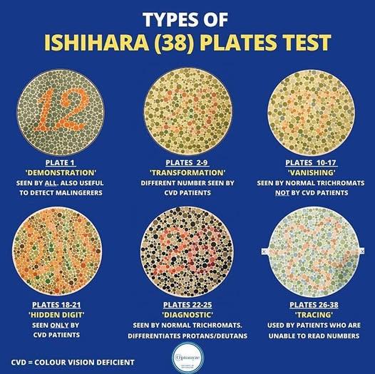
Ishihara test
- Normal trichromats failing IT have been termed “pigment farbanomaly” – fail printed test but not spectral tests
- IT remains the “gold standard” for rapid identification of congenital red-green CVD
- Individual plates are 85% sensitive and 95% specific for R/G CVD – test as a whole performs close to 100% sensitive and specific
- Ishihara is considered passed if the candidate makes less than 2 errors on plates 1-15 (24 plate test) and less than 4 errors on plates 1-21(38 plate test)
- If the candidate fails further investigation should be carried out – CAD at SACAA
Arrangement tests
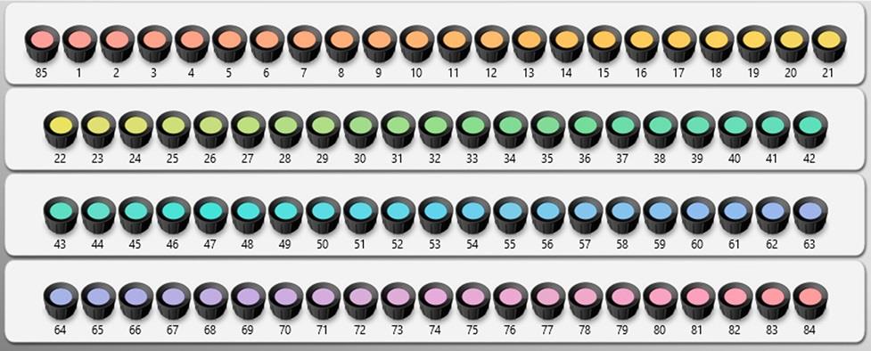
Arrangement tests involve asking observers to order a set of movable coloured objects according to either hue or saturation.
- Farnsworth Dichotomous Test for Colour Blindness (D- 15 test)
- The City University Test (CUT)
- The Farnsworth-Munsell 100 Hue Test
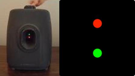
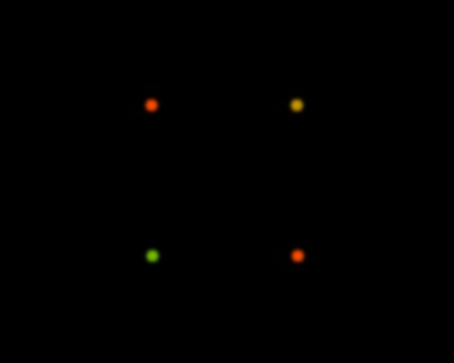
Lantern tests – Farnsworth lantern (FALANT)
- The lanterns are designed to mimic real-life situations, patients are presented with one or two lights at a distance and have to name them as soon as they see them
- Lanterns have seldom included blue stimuli, so tritan effects or defects are not likely to be considered
- Normal trichromats who failed Ishihara will all pass the lantern test
- A mixture of deutans and normal trichromats will pass the lantern test – some of the deutans may be colour unsafe
- All protans and some deutans will fail the latern test, though some may have sufficient chromatic sensitivity to be colour safe
Anomaloscope
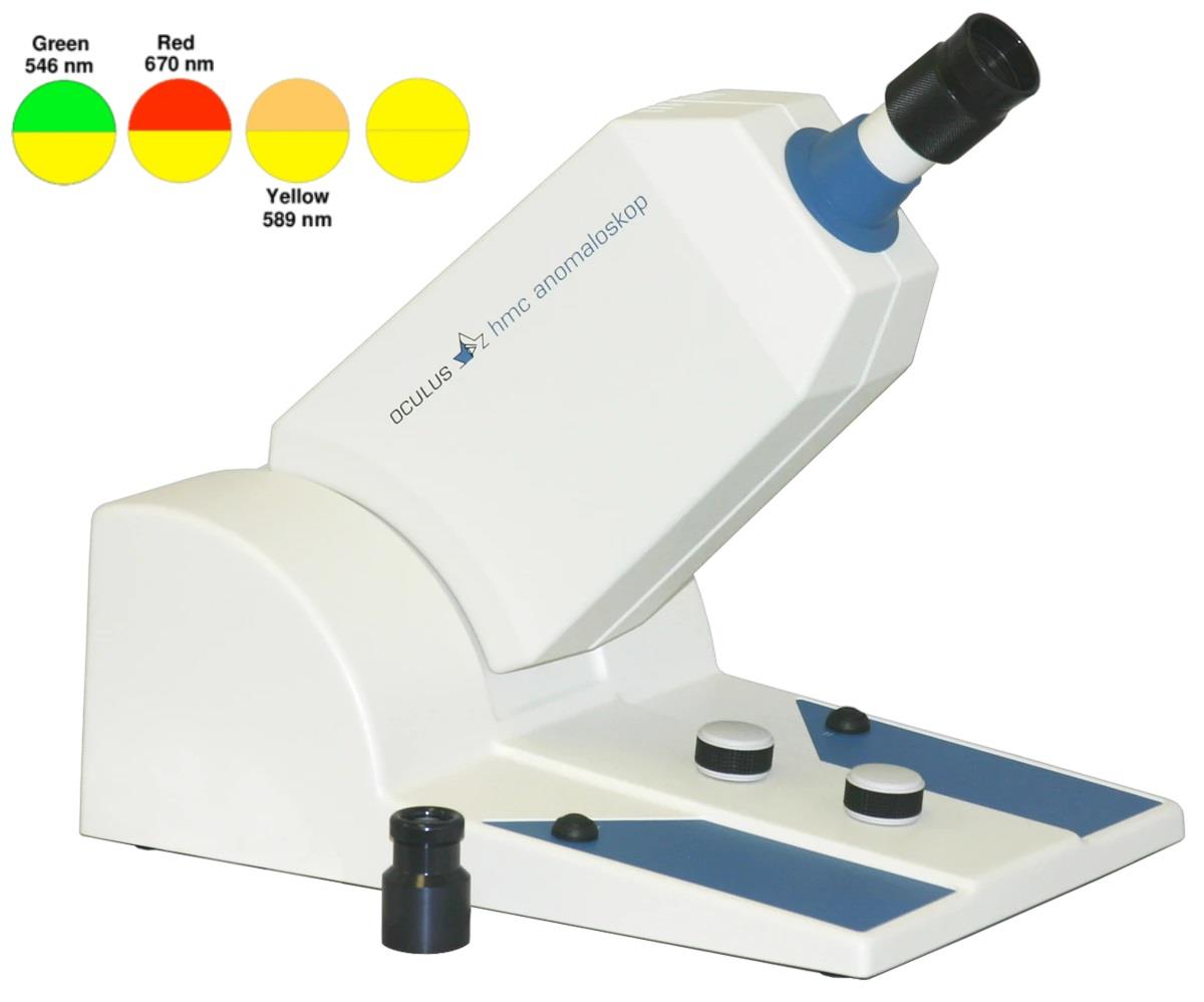
- The Nagel anomaloscope evaluates an individual’s Rayleigh matches, which are proportions of red and green light that need to be mixed to match the yellow light used as the test stimulus
- Tests for red-green deficiencies
- A vertically orientated bi-field composed of the top half (a mixture of red and green lights) and the bottom half (yellow light) is presented to a patient.
- The proportion of red and green lights in the mixture and the intensity of the yellow light are adjustable, and the patient is asked to control both the top and the bottom fields until a match is obtained
- Does not quantify well the degree of loss of colour sensitivity
- May not always detect colour deficiency
Computer based colour vision tests
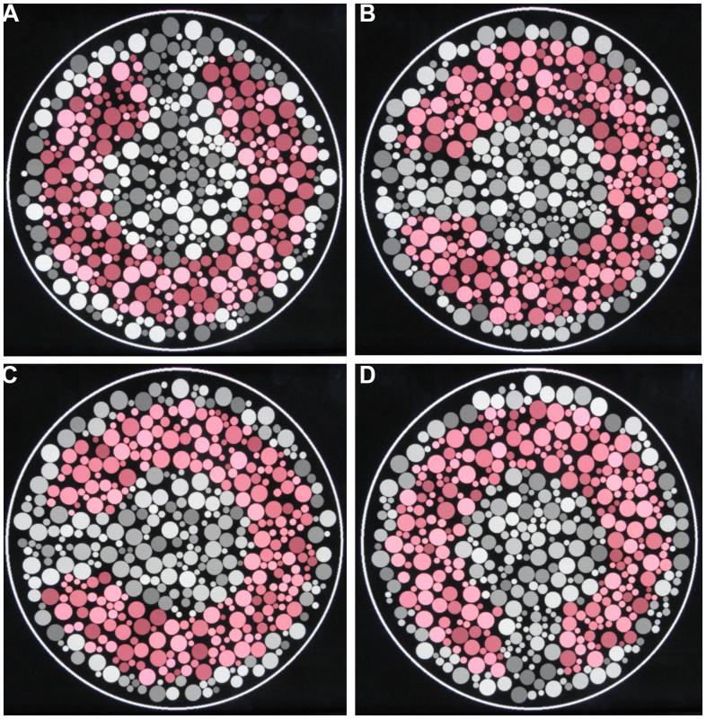
- The Cambridge Colour vision Test uses a computer version of pseudo-isochromatic plates and combines the Principles of Chibret and Stilling
- Each of the computer pseudo-isochromatic plates in the CCT contains a Landolt C of specific hue and luminance that is presented in four different orientations which is embedded in a background comprising circles of varying size, colour and luminance.
- The CCT stimuli backgrounds eliminate luminance or contour cues, and the figures (the test stimuli of Landolt letters) must be identified solely by their hues
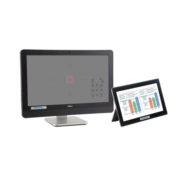
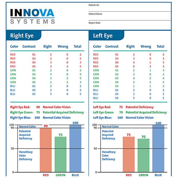
Rabin Cone Contrast Test (RCCT)
- The Rabin Cone Contrast Test (RCCT) is a specialized diagnostic tool used to assess the function of cone photoreceptor cells in the retina
- It measures an individual’s ability to perceive subtle differences in colour contrast across different wavelengths, providing a more detailed evaluation of colour vision than traditional tests.
- The RCCT is particularly useful in detecting and monitoring conditions like glaucoma, optic neuropathies, and other diseases affecting the visual system
- Pass – 75 or higher for red, green, and blue
- Each eye tested seperately
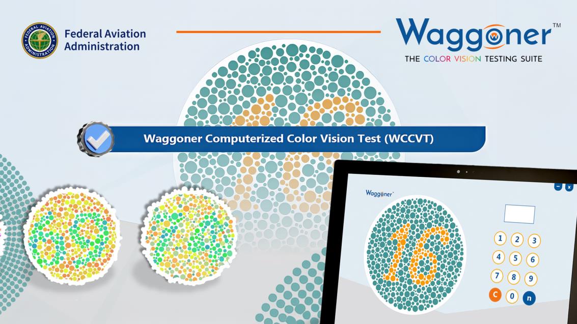
Waggoner Computerized Colour Vision Test (WCCVT)
- The Waggoner Computerized Colour Vision Test (WCCVT) is an all-in-one colour vision testing suite to satisfy anyone interested in testing for colour vision deficiencies ranging from school nurses to the U.S. Military
- Within the WCCVT, an individual can choose several different testing methods that includes screening, diagnostic, paediatric, and adult testing
- It has been coined one of three “precision” colour vision tests available today by the U.S. FAA
- It is a military-grade colour vision test that is accepted by the U.S. Navy, Army, and Coast Guard for all personnel and applicants, including pilots
- The diagnostic tests are perfect for identifying both genetic and acquired colour vision deficiencies and then providing the type and severity of the deficiency
Pass –
Generally 21/25 and tritan 10/12 OR individually
Tritan 10/12
Protan 20/32
Deutan 20/32
Problems with Current Secondary CVD Tests
- No test, including Ishihara* and anomaloscope**, assesses severity of colour vision loss
- Pass/fail variability (within-subject and inter-subject) is high*
- Lack of standardisation
- Correlation between outcomes of different tests is poor*
- Do not give reliable information about safe, minimum colour vision for flying
*Squire TJ, Rodriguez-Carmona M et al (2005). Color vision tests for aviation: comparison of the anomaloscope and three lantern types. Aviat. Space Environ. Med. 76, 421-429. 2 Barbur
**JL, Rodriguez-Carmona M et al (2008). A study of unusual Rayleigh matches in deutan deficiency. Visual Neuroscience 25, 507-516
Safety Critical Colour Discrimination Tasks in Aviation
Tasks with no redundancy (other cues):
- PAPI – Colour perception has to be accurate and fast
- Parking lights (much less hazardous)
Tasks with more redundancy or that are less demanding in terms of colour perception:
- Runway threshold, centre-line, lead-off, taxiway, stop way lights
- Navigation lights
- Rotating beacons on ground vehicles
- EFIS, maps, VASIS
CAD – Colour Assessment and Diagnosis Test and PAPI
- A team around Prof. Barbur from the Applied Vision Research Center in London was mandated to find the minimum colour vision requirements for modern flight crew, and a new Colour Assessment and Diagnosis test – CAD test – was developed
- The Precision Approach Path Indicator (PAPI) was indicated as the most important, safety-critical task that relies largely on colour vision in aviation
- The aim was to identify type and severity of colour vision deficiency which cause problems with the PAPI test and correlate those results to the CAD test results
- In principle, this approach should make it possible to recommend pass/fail limits based on the observer’s ability to carry out the most safety-critical and demanding PAPI task
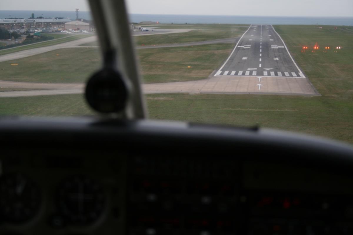
“White on white and you will fly all night”
“Red on red and you are dead”
Color Assessment and Diagnosis test
- The CAD test determines the threshold for perception of R/G and Y/B colour signals and quantifies the severity of colour vision using an internationally recognised colour system
- CAD has 100% specificity and 100% sensitivity for assessing whether an individual has normal colour vision (ie is a normal trichromat or not)
- CAD can quantify the severity of colour vision loss and it cannot be learnt
- Results are expressed in standard normal units, easy to understand, providing an immediate indication of the severity of the colour vision loss
- Useful in assessing changes in colour vision in subjects with diseases of the retina and optic nerve and to specify colour vision requirements in occupational environments
Barbur et al, Proc. Roy. Soc.B., 258, pp 327-334, 1994.
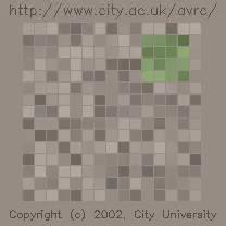
- Principal advantages of the CAD Test.
- 100% Sensitive and Specific
- Specific test for RG and YB
- Candidates that fail Ishihara and passed CAD:
36% Deutans
30% Protans
35% Overall
- Disadvantage
- Cost of test – only at academic institutions
- Subjects with minimum color blindness that does not exceed 6 SNU* units for deuteranomalous observers and 12 SNU units for protanomalous observers perform the PAPI test as well as normal trichromats
- >2 SNU for tritanomaly indicates an acquired color vision defect which should be investigated and is an automatic failure
- These findings were adopted as pass/fail limits for pilots
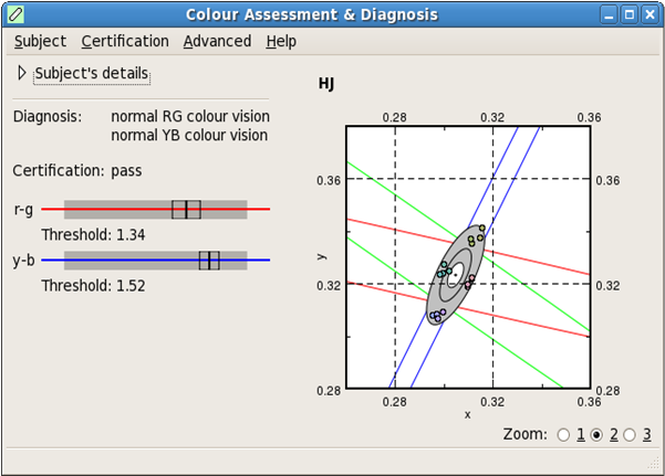
*SNU – standard normal unit – red-green threshold 2SNU – twice as strong colour signal needed compared to average CAD observer
Why such stringent color standards?
Color is superior to other coding modalities, such as size, shape, frequency, brightness, number, orientation, position:
Shorter reaction time (50-90% faster)
Reduced error rate (200% more accurate)
The benefit of color increase with increasing display complexity and task difficulty
Challenges facing the color-defective pilot
- Complex color-based environment
- Extreme demands on the visual system
- Must be closer to the target to discriminate it based on color information
- Take longer to interpret color-based information
FedEx 1478, 2002 struck trees on final approach to Tallahassee- first officer (flying at the time) color deficient – could not interpret the PAPI indicator – contributing to the accident
SACAA standard CVD protocol – DAMES p.216
- Applicants who fail Ishihara and CAD shall be assessed as unfit
- A class 2 medical certificate may be issued if medical conclusion indicates that the applicant has a colour perception defect which is compatible with the safe exercise of the privileges of the license, provided the certificate is endorsed with the following limitations and the following conditions are met:
- “For private pilot license privileges only”;
- Not valid for night flying;
- Not valid for IFR flying or flying of EFIS-equipped aircraft where the EFIS is the primary flight instrument;
- The applicant meets the visual criteria for a Class II medical certificate;
- The applicant shall submit a satisfactory report from an ophthalmologist every two years if the if < 40 years of age and every year if > 40 years of age.
Operational colour vision test (OCVT) & Medical practical flight test (PMFT)
- The PMFT shall be conducted in a Level C or D simulator, or such lessor device as determined by the Director
- Conducted by a panel of specialists appointed by the Director and will be coordinated by authorised officers (medical assessors) of the SACAA;
- Panel shall comprise of the following: A representative authorised officer from the CAA; A designated aviation medical examiner, preferably with experience in flying; An ophthalmologist; A designated flight examiner as determined by the Director;
- The procedure for the medical practical flight test shall be approved by the Director
- Applicants must demonstrate the following: Must read and correctly interpret in a timely manner aviation instruments or displays, particularly those with coloured limitation marks; Must read and interpret coloured instrument panel lights, especially marker beacon lights, warning or caution lights, weather displays, etc.;
- Must recognise terrain and obstructions in a timely manner, have the applicant select several emergency landing fields, preferably under marginal conditions, and describe the surface; Must visually identify in a timely manner the location, colour and significance of aeronautical lights.
- Applicants will be afforded a single opportunity for a medical practical flight test.
Considerations for applicants with Class I who fail a CAD and pass the OCVT and PMFT tests
To fly as CPL in a multi-crew environment by day and night as a deuteranope with the following restrictions:
- The holder does not meet the ICAO medical standard as per Annex 1 and is therefore restricted to fly within the South African borders on a South African registered aircraft only
- Applicants who fail the CAD will not qualify for Air Transport Pilot Licence operations.
- Annual ophthalmological assessment will be required to determine any refractory, visual field or lens translucency change every two years if < 40 years and annually if > 40 years.
- The applicant must inform his/her employer and cockpit crew members of his/her red-green colour deficiency.
- The holder is restricted to a cabin altitude of maximum 8000 ft AMSL at night or during IFR conditions.
- The holder may not perform any CAT II approaches.
- A minimum required flight hours as prescribed in SA-CARS/CATS Part 61 will be applied before allowing the applicant as PIC with CPL.
- The decision and restrictions will be reviewed, should there be a change in the applicant’s condition or new evidence becomes available regarding deuteranopia and flight safety
Common ophthalmic conditions in aviation
- Conjunctivitis
- Cases should be assessed individually. Viral conjunctivitis is associated with sore throat and lymphadenopathy and is infectious. Conjunctivitis often requires topical medication (e.g. antibiotic drops) and can impact on certification when purulent secretions are copious or when drop administration is very frequent. Please note that topical eye ointments may cause reduced vision for a period after application.
- Minor eyelid infections
- Blepharitis, meibomian cyst (chalazion) or stye (hordeolum) do not normally impact on certification unless they cause discomfort or reduction in vision (e.g. due to mechanical ptosis or induced astigmatism). Topical treatment with warm compresses and topical medication, if required, can present practical problems with a busy flying schedule and so cases should be assessed individually.
- Keratitis
- Certificate holders should be assessed as unfit on diagnosis. Restoration of fit status can be considered once the condition has resolved and the applicant has stopped all medication, or occasionally when it is accepted that the condition requires low dose maintenance topical therapy.
- Anterior Uveitis
- Certificate holders should be assessed as unfit on diagnosis. Restoration of fit status can be considered once the condition has resolved and the applicant has stopped all medication, or occasionally when it is accepted that the condition requires low dose maintenance topical therapy. Consideration should be given to underlying causes (such as ankylosing spondylitis), especially if the uveitis is recurrent.
- Posterior Uveitis
- This is associated with underlying disease (inflammatory bowel disease, sarcoidosis, etc). Certificate holders should be assessed as unfit on diagnosis and a formal consultant ophthalmologist report is required (see CAA’s guidance on ophthalmic reports) and should include the results of all systemic investigations. A return to fit status should be considered bearing in mind visual function, medication and identification and control of any underlying cause.
- Trauma
- Eye injuries sufficiently severe to require medical attention will require a formal ophthalmological report. Cases should be assessed individually and suspension of the validity of the medical certificate may be required. Class 1 and 3 holders may be required to undertake assessment with a consultant ophthalmologist.
- Cataract
- This is compatible with fit status provided that: visual standards are met; and there are no symptoms of glare, halos etc.
- Retinal Detachment
- Certificate holders should be assessed as unfit on diagnosis. Consultant ophthalmologist reports will be required. Restoration of fitness status is possible provided that the effect on visual function is such that the visual standards are met. Peripheral retinal tears treated successfully with laser can be considered for restoration of fit status following recovery from successful treatment. In complex retinal detachment resulting in loss of peripheral field, certification should be discussed with a CAA Medical Assessor.
- Central Serous Retinopathy
- Certificate holders should be assessed as unfit on diagnosis. Restoration of fitness status is possible when the condition has resolved or when no further improvement to vision is expected provided that the visual standards are met. If necessary, please also refer to the substandard vision in one eye guidance.
- Acquired Disorders of the Macula
- Certificate holders should be assessed as unfit on diagnosis as macular disorders can cause significant distortion (metamorphopsia) without necessarily reducing vision to below acceptable limits. Fitness may be reconsidered when ophthalmological reports are received. Restoration of fitness will be considered on an individual case basis as there is such a wide spectrum of macular disease severity. If necessary, please refer to the substandard vision in one eye guidance.
- Glaucoma and Ocular Hypertension
- The initial diagnosis should be reported to the CAA/AME and visual function assessed. In uncomplicated cases routine follow up reports (including visual fields) can be taken to the AME at each medical. In severe cases, please refer to the visual field and substandard vision policies, as well as the guidance on eye surgery. Applicants with any form of open or closed angle glaucoma may be considered for certification provided that the consultant ophthalmology reports confirm an acceptably low rate of recurrence.
- Vascular occlusions Including – retinal artery occlusion, ischemic optic neuropathy, amaurosis fugax, and retinal vein occlusion.
- Pilots with arterial vascular disease affecting the eye should be made unfit. The subsequent aeromedical fitness assessment needs to take into account both the effect on visual function and the cardiovascular incapacitation risk.
- It is important to identify disease due to emboli from the left side heart and carotids, as this carries a higher cardiovascular risk. Infective endocarditis and the systemic vasculitis’s, including giant cell (temporal) arteritis and thrombophilia must all be excluded, as these conditions have their own treatment protocols and aeromedical implications.
- Arterial vascular disease affecting the eye is usually associated with an increased cardiovascular mortality. Cardiovascular risk factors must be identified and managed before re-certification.
- Pilots with retinal vein occlusions (RVO) should be declared unfit. The subsequent aeromedical fitness assessment needs to take into account both the effect on visual function and the cardiovascular incapacitation risk. RVO reduces visual acuity and field of vision in the affected eye, sometimes permanently.
- RVO is usually associated with an increased cardiovascular mortality. High blood pressure is a cardinal risk factor for RVO and satisfactory blood pressure control is therefore essential before re-certification.
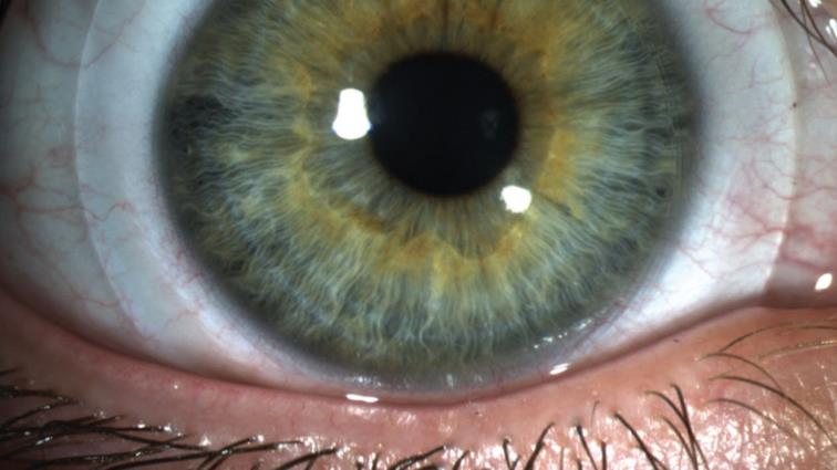
Keratoconus
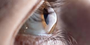
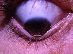
- “Keratoconus is a bilateral, and asymmetric corneal degeneration characterised by localized corneal thinning which leads to protrusion of the thinned cornea”
- Thinning normally occurs in the inferior-temporal and central cornea (can be anywhere on the cornea)
- Ectasia causes high myopia, irregular astigmatism, and reduced visual acuity
Diagnosis and Classification
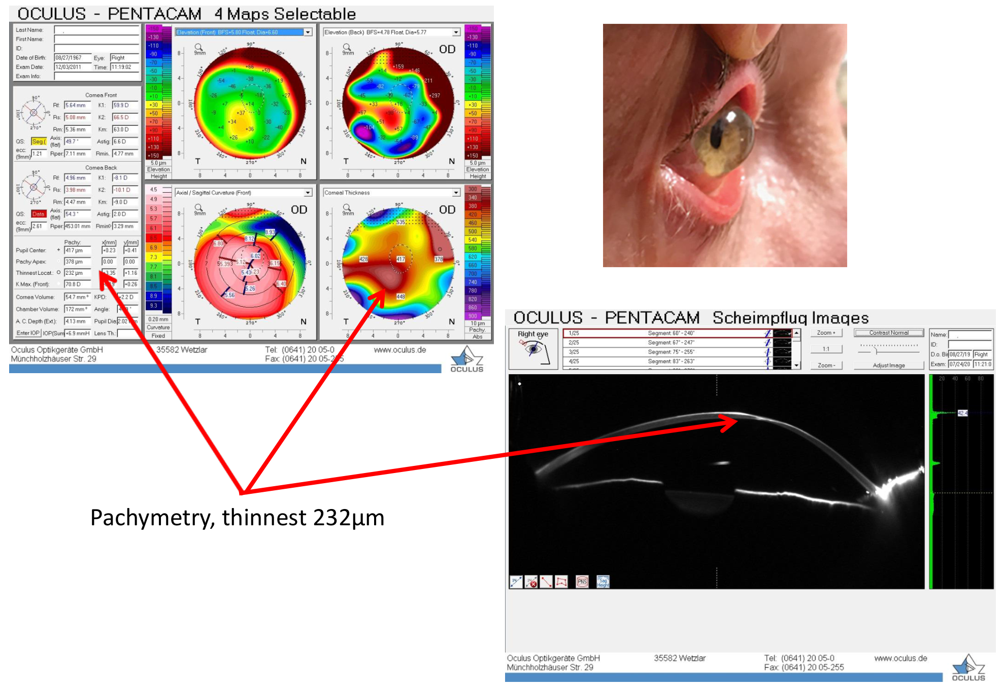
- Both changes on the posterior corneal surface and alteration in the corneal thickness progression are necessary to diagnose early stages of KC
- Tomography (Scheimflug and OCT) and slit-lamp examination are currently the best and most widely available tests to diagnose early KC
- A “gold standard” classification for KC is not currently available
- The often used Amsler-Krumeich classification system is limited, additional information including visual performance (BVA) correlated with corneal topometric and tomographic parameters may improve the classification and diagnosis of KC
Progression
- Ectasia progression is defined by a consistent change in at least 2 of the following parameters
- Steepening of the anterior corneal surface
- Steepening of the posterior corneal surface
- Thinning and or an increase in the rate of corneal thickness change from the periphery to the thinnest point
- Although VA often decreases with progression, changes in uncorrected and corrected VA is not required to document progression
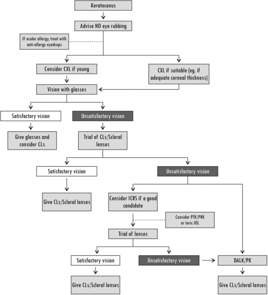
Non-surgical treatment options (Gomes, Tan et al. 2015)

Management of KC
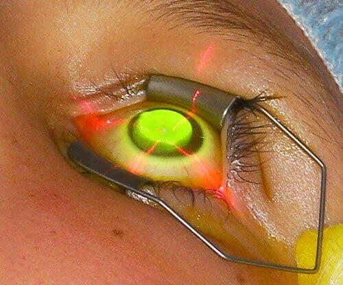
- Most important goals of management are halting progression and visual rehabilitation – corneal crosslinking
- In cases of allergy, atopy or eye rubbing, patients must be treated with topical anti-allergic medication, topical lubricants and counselled not to rub their eyes
- There is no direct relationship between KC and dry eye
- Contact lenses does not slow or halt progression of corneal ectasia
- Pregnancy could contribute to accelerated progression of ectasia
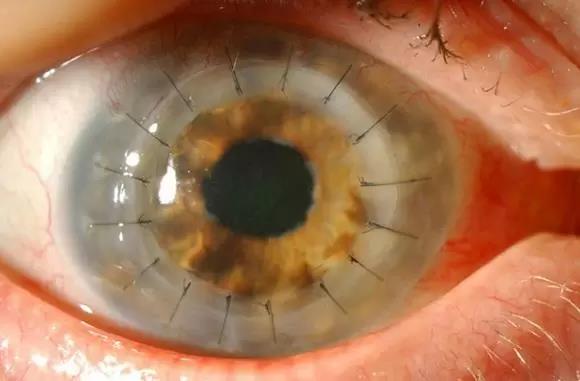
- In acute hydrops, nonsurgical management should be attempted before keratoplasty
Ophthalmic drugs in aviation
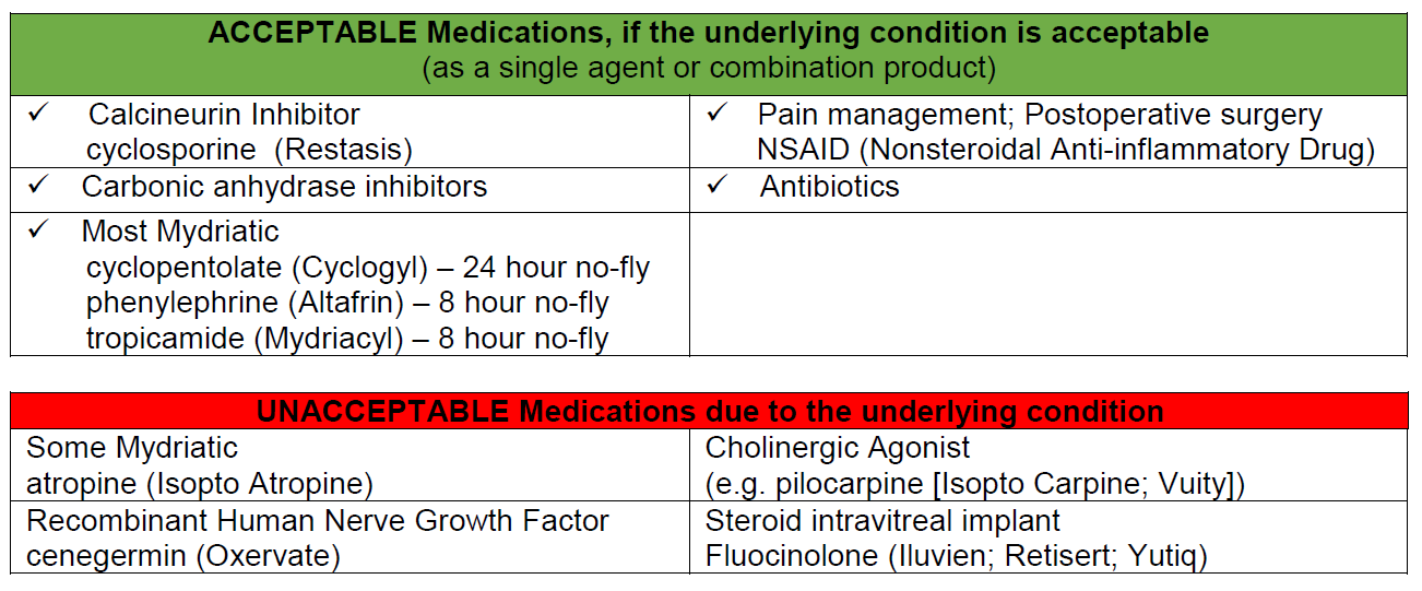
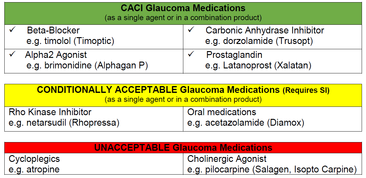
Glaucoma drugs in aviation
Presbyopic Ophthalmic Drugs in Aviation
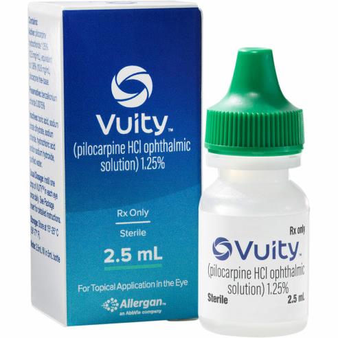
- Recently cholinergic agonists (parasympathomimetic) drops have become available to treat presbyopia
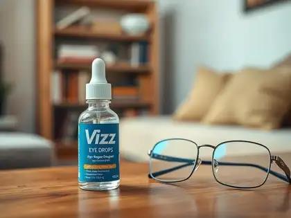
- The active ingredients include pilocarpine (Vuity) and aceclidine (Vizz) which increases depth of focus by inducing miosis of the pupil
- Effects are said to last up to 6 hours and adverse effects were rare but include retinal detachment and accommodative spasm as well as headaches.
- For obvious reasons these are not acceptable drugs in aviation
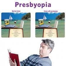
Presbyopic Pilots
- Add power for flying is different than for desk work
- Pilots may need a different pair of specs for flying than general work
- Lenses must produce the minimum amount of distortion and require minimal adaptation time
- Best choice flat top bifocals, inferior temporal area of the lens helps during the landing flare
- Multifocals have distortion in this area, many pilots fly successfully with these lenses, one type of multifocal does not suit all pilots in all aircraft
- Hypoxia may cause problems with perceived multifocal lens distortion
- Bifocal height is crucial
- Set the bifocal segment just above the top instruments, and just below the glare shield
- Helmets, headsets affect the fit of the frame
- Pilots lean their head back during the landing flare, bifocal can interfere leading to early flare as runway appears larger (closer)
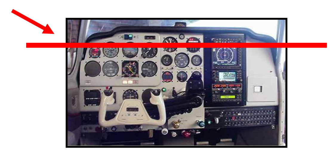
Increased minus or plus spectacle power
- Increase in minus power leads to smaller images and object appear further away
- May cause
- Late flare on landing
- Hard landing with possible damage to landing gear/airframe
- Increase in plus power leads to larger images and object appear closer
- May cause
- Early flare on landing
- Damaged landing gear/airframe

Sunglasses
- In general, not less than 15% light transmittance, no color distortion, no Polaroid
- Reduce visual fatigue by reducing exposure to glare
- Good idea to use during the day before night flying
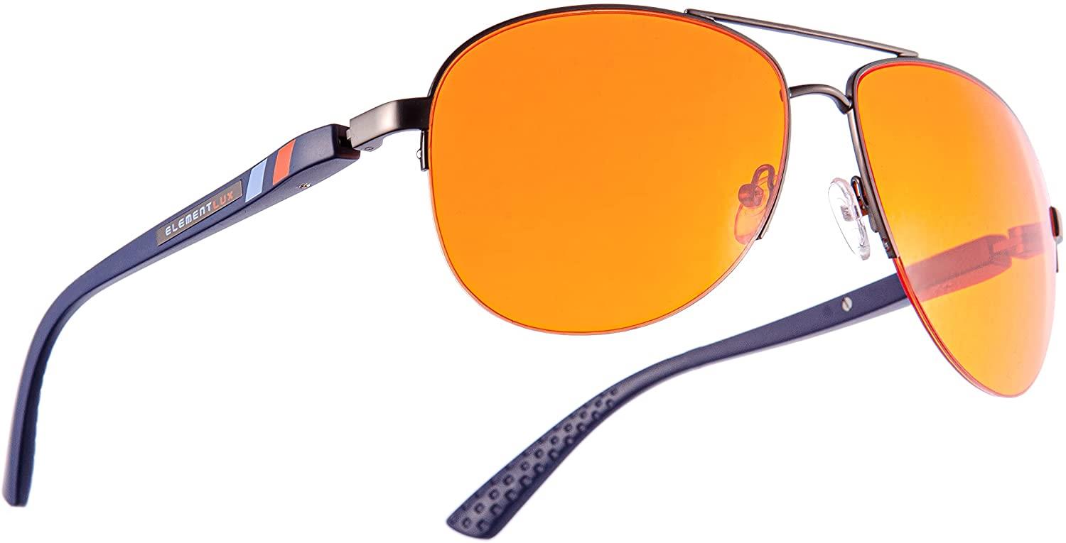
Selective filters
Blue blockers and yellow lenses interfere with color perception, therefore not allowed
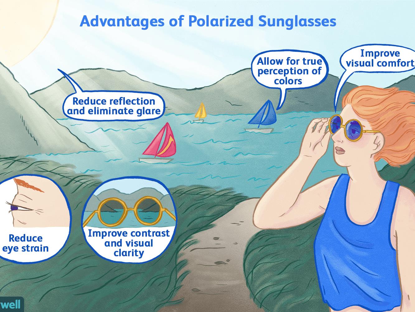
Polaroid sunglasses
- Excellent for glare off flat surfaces such as water
- Problem in aircraft due to plastic windshields & canopies causing “dark areas & blind spots”
- Liquid crystal displays can be problematic
- Not good choice for pilots
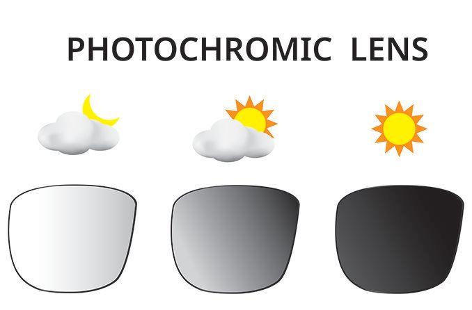
Photochromics
- Less UV light in cockpit due to polycarbonate wind. screen, so lens not as dark
- Temperature dependent
- Delay to lighten can cause problems when flying into cloud
Standard neutral density filters
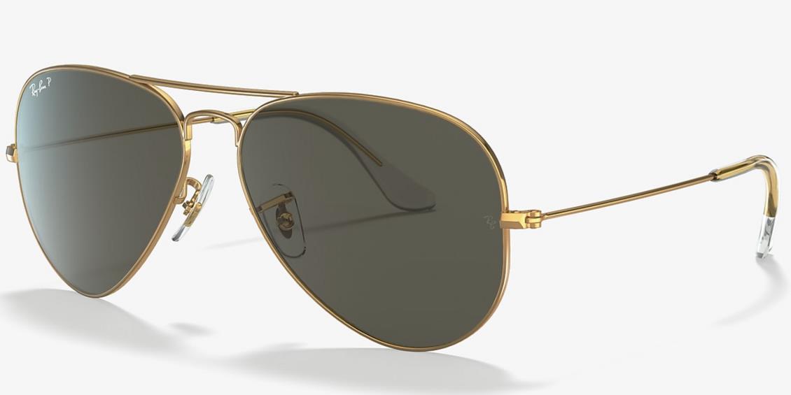
- Relatively flat transmission curves
- No-color distortion
- Only sunglass lens authorized for air force aircrew (N-15 or G-15)
SACAA Ophthalmology Report Template
- Aviator or applicants name, licence class and age
- History
Systemic and ophthalmic - Vitals
Visual acuity with and without correction
Pupil reactions
Intra ocular pressure and method used to measure it (GAT, NCT and RBT) - Refractive status
- Eye movement and alignment – strabismus or phoria and size of deviation if any
- Anterior segment evaluation – lids, adnexa, conjunctiva, cornea, anterior chamber and crystalline lens
Corneal topography and tomography scans if available - Posterior segment evaluation – ocular media, macula, disc, mid periphery and periphery
Fundus photos, ocular coherence tomography, and visual field tests if available - Diagnosis, prognosis and treatment plan
Thank You
dirk@dirkbooysen.co.za