
ISCLS Italy 2024
dirk@dirkbooysen.co.za
Case report
- 28 year old Caucasian male keratoconus patient
- Reports painful right eye, mild redness, white cornea and poor vision
- Cannot wear his lens at all
- Prior to this incident had 20/30 vision with RGP
- K-readings were 5.86/5.56 mm
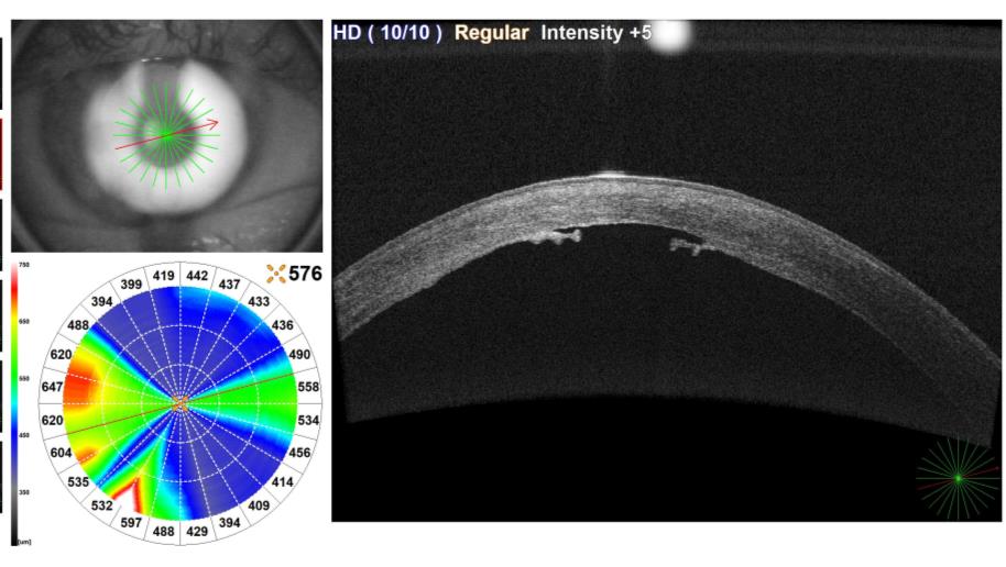
- Pachymetry 219 µm, prior to hydrops
- IOP 8mmHg
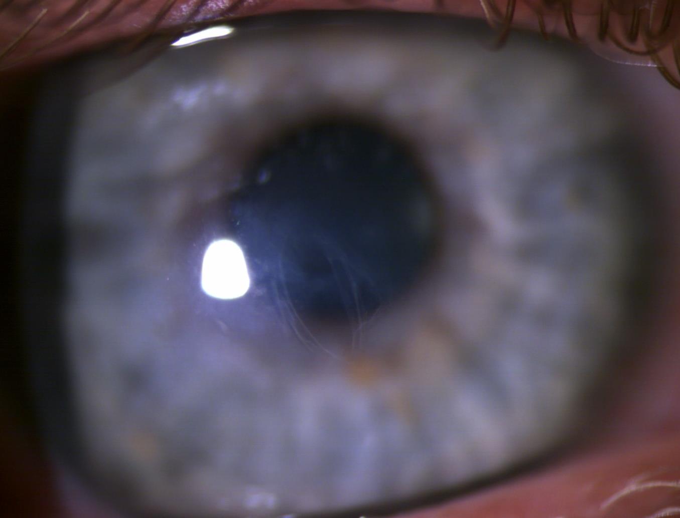
- Central scarring present
- RGP lens 5.50BC/5.50OZ/9.10OD/-20.50D Aspheric e-value 1.0
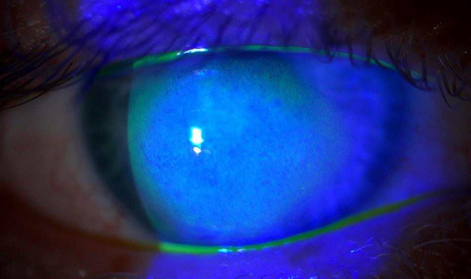
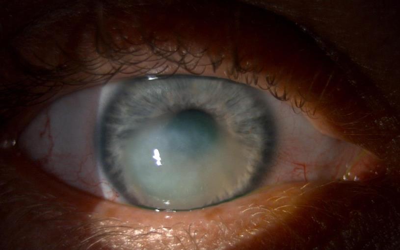
Diagnosis – Acute Hydrops
- Prevalence: In keratoconus patients is 2.6 to 2.8%.
- Risk factors: Vernal keratoconjunctivitis, atopic dermatitis, high keratometry, male gender, and eye rubbing.
- Resolution: Corneal oedema typically resolve within months (≥100 days).
- Complications: Corneal perforation, infectious keratitis, and corneal vascularization may occur
Mechanism
- CH occurs due to the torn DM which causes the rolling of the edges, a gap is created so that the aqueous from the anterior chamber seeps into the corneal stroma.
- Continuous accumulation of the aqueous leads to the separation of the collagen lamellae and the formation of large fluid-filled stromal pockets.
- Endothelium grows over the defect causing a partial seal so that the seepage is prevented with subsequent resolution of stromal oedema
- Keratoplasty should be avoided in the acute phase
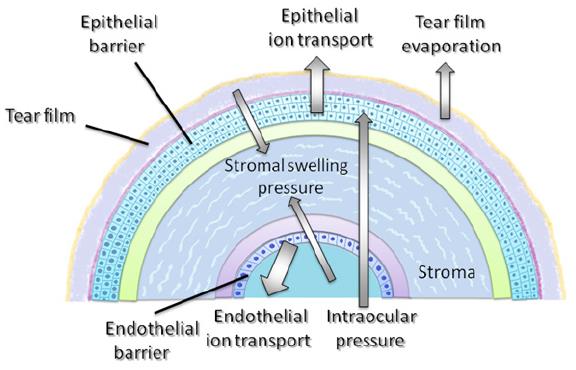
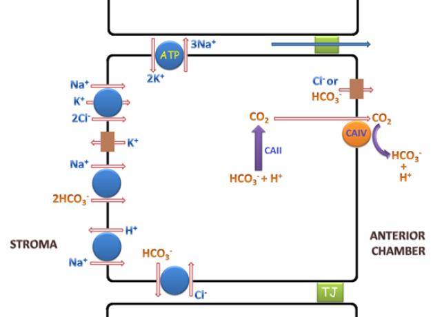
- Corneal transparency is maintained owing to the balance between the pressures that cause either input of the fluid into stroma or output from it
- While stromal swelling pressure and intraocular pressure draw water into the stroma, endothelial and epithelial ion transports (membrane-bound Na+/K+ ATPase and the intracellular carbonic anhydrase pathways) ensure the dehydration of the cornea
- Tear film evaporation is another factor that contributes to this dehydration state
- Additionally, there are two barriers that prevent the movement of the fluid in both directions (anterior and posterior): the epithelial barrier and the endothelial barrier
- It is estimated that the endothelial fluid pump removes around 2.5ml of fluid from the cornea each day
Medical treatment of Acute Hydrops
If you can quickly resolve oedema in acute hydrops cases, you’ll reduce the need for a transplant
- Topical hyperosmotic: Can reduce corneal oedema (albeit slowly), and improve acuity
- Topical steroids: Manage corneal oedema and reduce risk for neo-vascularization. Usually twice-daily dose
- Since steroids have the potential to hinder corneal healing or cause corneal perforation, initiating steroid treatment at onset, or even once DM membrane heals, is controversial
- Cycloplegic agent: Twice per day (more if pain persists), reduce ocular pain from a secondary uveitis and relax ciliary muscle
- NSAID’s: Another option for pain relief include oral or topical nonsteroidal anti-inflammatory agents
- Bandage contact lens: Pain relief but keep in mind that depending on severity of KC the bandage lenses can flute and decentre
- Topical antibiotics: If epithelial defects are present, a prophylactic antibiotic should be prescribed to decrease the potential for infection
- Oral doxycycline and vitamin C: 10mg to 50mg twice per day and vitamin C ,1,000mg per day—typically dosed early in the course of acute hydrops—can be beneficial
- Doxycycline may also reduce matrix metalloproteinases, which promotes corneal healing
- Vitamin C has a role in the extracellular matrix and corneal composition, and can decrease opacification and neovascularization
- Amniotic membrane transplants and copious preservative-free artificial tears
Surgical options for Acute Hydrops
Healing under conservative therapy takes an average of over 100 days
- Air/gas tamponade: If the DM is detached without tension, a simple injection of air or gas (20% sulphur hexafluoride (SF6) or 14% perfluoropropane (C3F8), into the anterior chamber to mechanically close the opening in DM can lead to almost immediate deswelling of the cornea
- Predecemetal sutures: If the DM is under tension, predescemetal sutures combined with a gas injection into the anterior chamber can flatten the cornea and reattach the DM.
- Mini-Descemet membrane endothelial keratoplasty: Mini-DMEK allows for suture less closure of the DM defect by transplantation of a small (< 5 mm) graft.
- Mini-DMEK can then lead to permanent healing, but in contrast to simple corneal sutures, it is usually performed under general anaesthesia and by aid of intraoperative optical coherence tomography
- Surgical therapy offers rapid healing and good results making it an option for most acute hydrops patients
- For maximum effect, these procedures must be per-formed typically within days after the initial onset of symptoms
- Surgery may improve healing time but do not impact visual acuity outcomes, making the procedures controversial due to potential adverse events
Novel treatment plan
With regime the recovery time decreases from months to weeks
- Anti-glaucoma drugs: Either prostaglandin analogues such as Xalatan (latanoprost 0.005%) or combination drugs such as Xalacom (latanoprost 0.005% /timolol 0.5%) or Duotrav (travoprost 0.004%/timolol 0.5%) one drop at night
- These drugs reduce aqueous production and increase aqueous outflow lowering IOP
- Reduced IOP lessens the hydrodynamic forces on the posterior cornea
- Do not use a topical CAI as it influences the endothelial pump – blocks isoenzyme CA II, decreasing HCO3– production resulting in diminished pump activity, leading to stromal swelling
- Topical antibiotic: Exocin – 0.3% ofloxacin qid, to prevent secondary infection if epithelium is compromised
- Topical steroid: FML – 0.1% fluorometholone suspension qid, only if needed
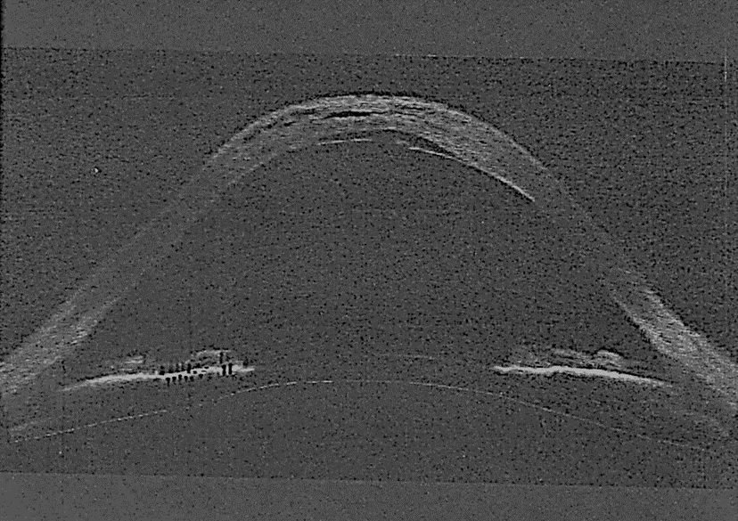
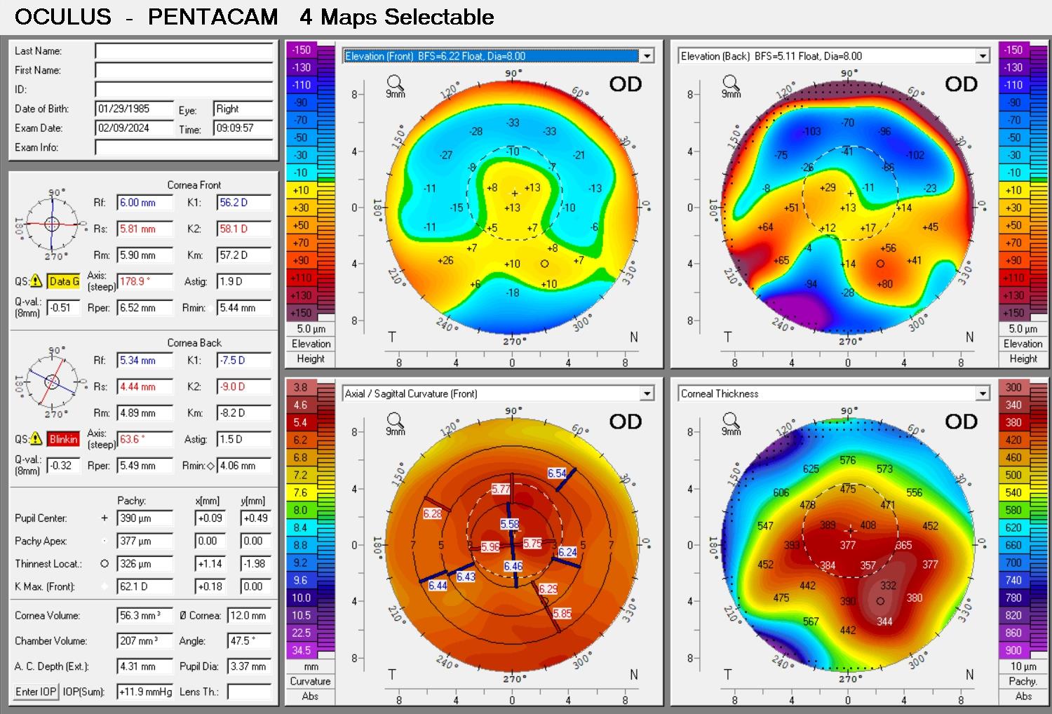
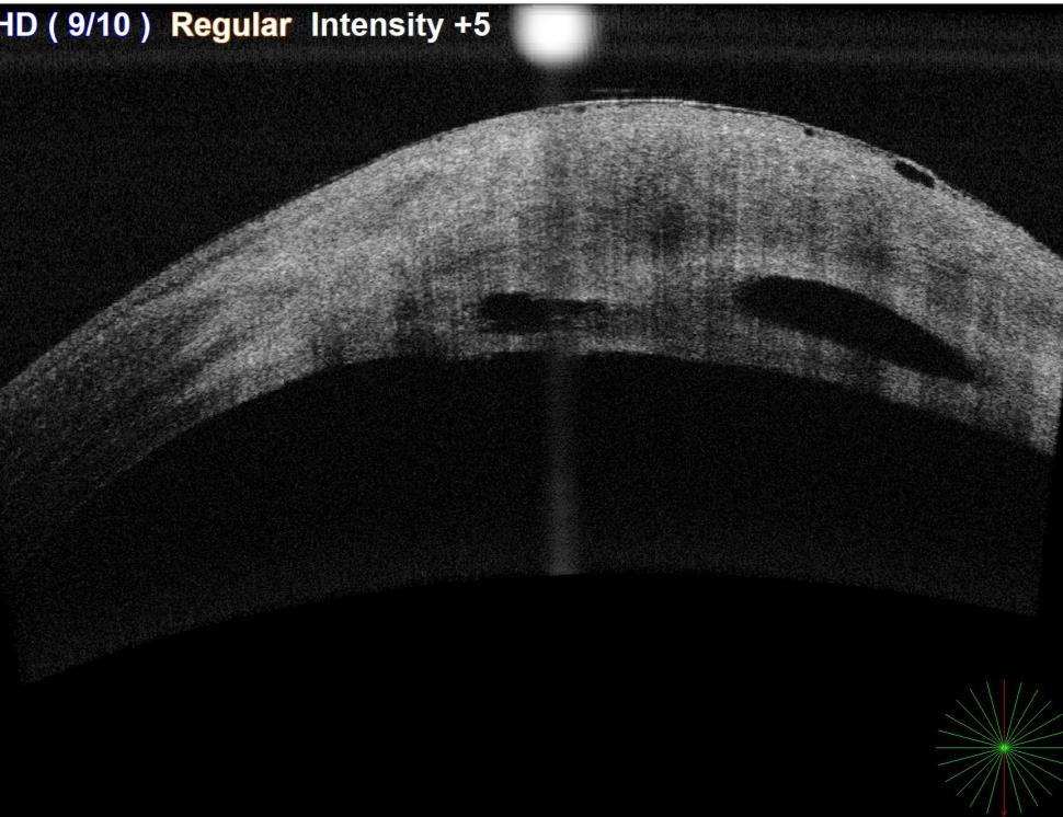
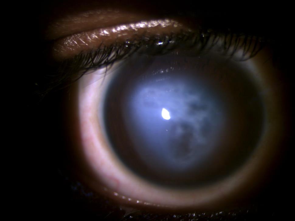
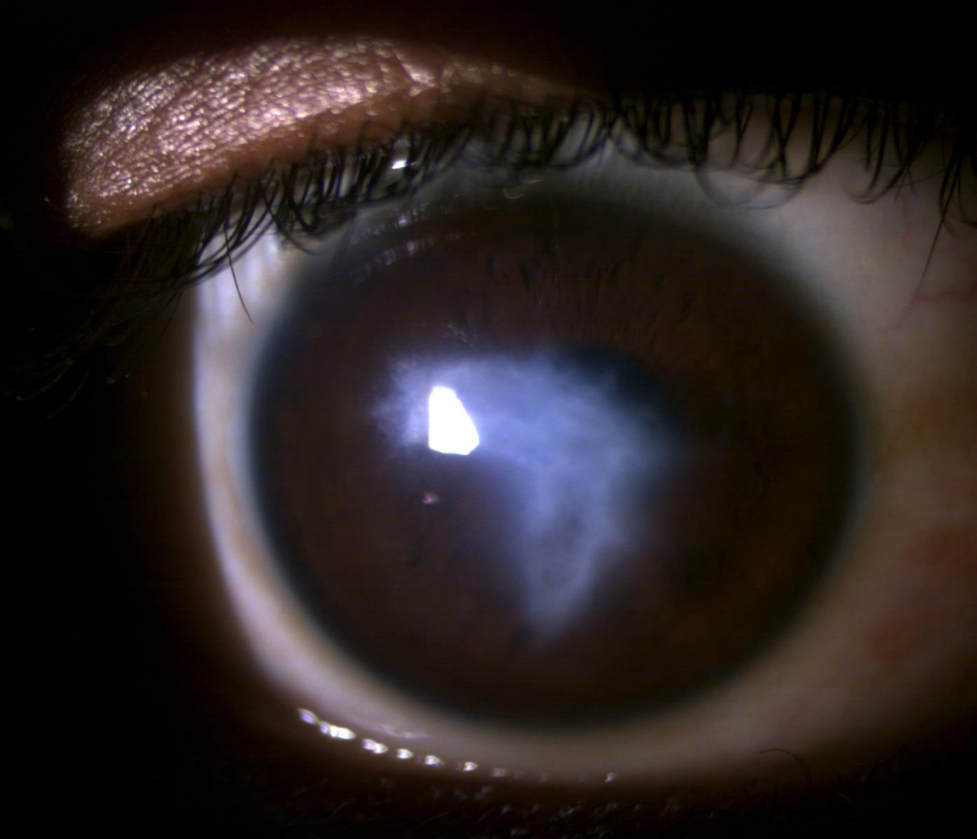
Pentacam®, photo and OCT at last visit
Final word
- We should strive to minimize patient symptoms and avoid devastating long-term complications with appropriate, safe and uncomplicated medical management
- Diagnostic imaging equipment can be helpful not only with initial diagnosis, but also in monitoring resolution
- Corneal hydrops is relatively rare*, advances in detection and treatment of keratoconus with corneal crosslinking, may reduce or eliminate it in the future
* If you work with keratoconus this statement is not entirely accurate
References
- Godefrooij D, de Wit G, Uiterwaal C, et al. Age-specific incidence and prevalence of keratoconus: a nationwide registration study. Am J Ophthalmol. 2017;175(3):169-72.
- Gokhale N. Epidemiology of Keratoconus. Indian J Oph-thalmol. 2013;61(8):382-3.
- Barsam A, Petrushlin H, Brennan M, et al. Acute corneal hydrops in keratoconus: a national prospective study of inci-dence and management. Eye (Lond). 2015;29(4):469-74.
- Gokul A, Krishnan T, Emanuel P, et al. Persisting extreme acute corneal hydrops with a giant intrastromal cleft secondary to keratoconus. Clin Exp Optom. 2015;98(5):483-6.
- Thimons J. Managing acute corneal hydrops in keratoconus. Rev Cornea & Cont Lens. 2015;152(3):32-3.
- Fuentes E, Sandali O, El Sanharawi M, et al. Anatomic predictive factors of acute corneal hydrops in keratoconus: an optical coherence tomography study. Ophthalmol. 2015;122(8):1653-9.
- Cho Y, Yoo W, Kim S, et al. Efficacy of systemic vitamin c supplementation in reducing corneal opacity resulting from infectious keratitis. Medicine (Balt). 2014;93(23):1-8.
- Perry H, Hodes L, Seedor J, et al. Effect of doxycycline hyclate on corneal epithelial wound healing in the rabbit alkali burn model. Cornea. 1993;12(5):379-82.
- Maharana P, Sharma N, Vajpayee R. Acute corneal hydrops in keratoconus. Indian J Ophthalmol. 2013;61(8):461-4.
- Shaw J. Acute hydrops: rethinking treatment. EyeNet Magazine. www.aao.org/eyenet/article/acute-hydrops-rethinking-treatment. June 2012. Accessed October 15, 2018.