
The Scleral Lens Education Society: Case Report 4
Dirk J Booysen
Dip. Optom FOA(SA), MCOptom(UK), TMOD(PCO), CAS(NECO), DOptom(Aston)
248 Voortrekker Road
Monument
Krugersdorp
Republic of South Africa
Abstract
Corneal injury either by trauma, surgery or infection often results in severe scarring and generalised or localised leukoma or white vascularized corneal opacities involving the cornea. The affected eye often squints due to loss of sensory input and the scarred cornea is extremely irregular resulting in high irregular astigmatic refractive errors and paracentral fixation. Using a rigid gas-permeable mini-scleral contact lens can improve visual outcome significantly.
This case report deals with the fitting of a mini-scleral rigid gas permeable contact lens in a patient with a localized corneal leukoma adherens to improve visual acuity.
Key Words
Rigid Gas-Permeable Semi-scleral gas permeable lens, leukoma, leukoma adherens, wall eye, and neonatal conjunctivitis
Introduction
Neonatal conjunctivitis or ophthalmia neonatorum is defined as conjunctival inflammation developing within the first four weeks of life 1,2. The infectious agent (bacteria, virus, chlamydia) can cause severe localised infection of the eye including the cornea as well as a potentially serious systemic infection 1 2. Precise identification of the cause of the infection is therefore essential. Although the infection is often transmitted from the mother to infant during delivery, a number of factors contribute to the cause of neonatal conjunctivitis. They include 1:
- Organisms harboured in the mothers birth canal
- Treatment of maternal infections during pregnancy
- Duration and site of exposure of the infant to infectious organisms
- Adequacy of ocular prophylaxis immediately after birth
- Susceptibility of the infants eye to infection
- Amount of ocular trauma during delivery
The most likely causes of neonatal conjunctivitis include 1,2:
- Chlamydia trachomatis and Neisseria gonorrhoeae (more common in third world countries), maternal chlamydia infection rates in South Africa vary between 4.7 to 13% amongst antenatal clinic attendees 3
- Herpes simplex virus typically HSV-2
- Staphylococci and streptococci, Haemophilus influenza and various gram negative organisms
- Topical preparations used as prophylaxis against infection
The most likely cause of the neonatal conjunctivitis/keratitis in this case was probably Chlamydia trachomatis or Neisseria gonorrhoea, both common causes of neonatal infections in rural South Africa. Although chlamydial infections are often mild and self-limiting severe cases may occur that result in conjunctival and corneal scarring 1. The gram negative N.gonorrhoea can penetrate the intact corneal epithelium and result in a hyper acute conjunctivitis or keratitis with marked eye lid oedema, profound chemosis, and excessive purulent discharge. Corneal ulceration with perforation can occur if the infection is not treated adequately 1.
Case Report
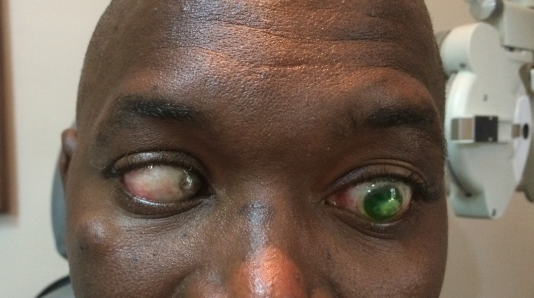
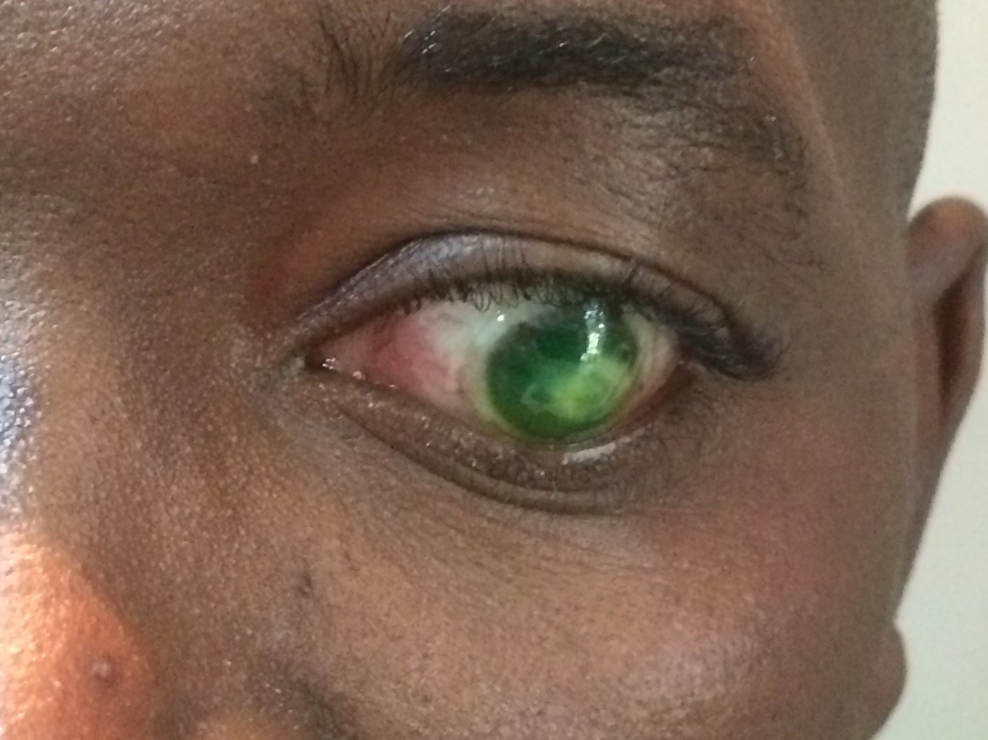
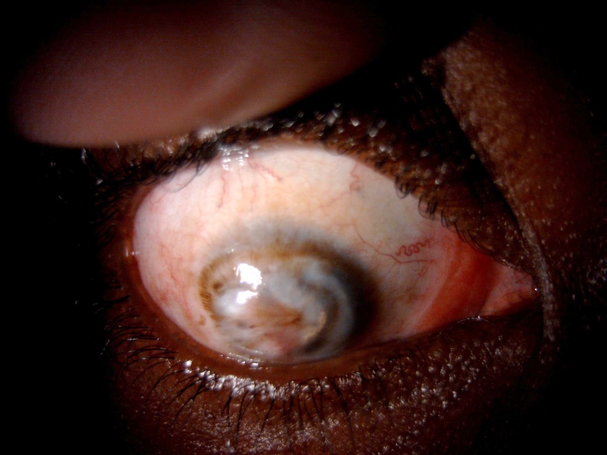
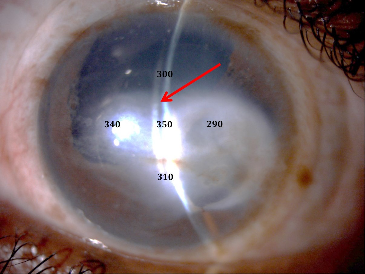
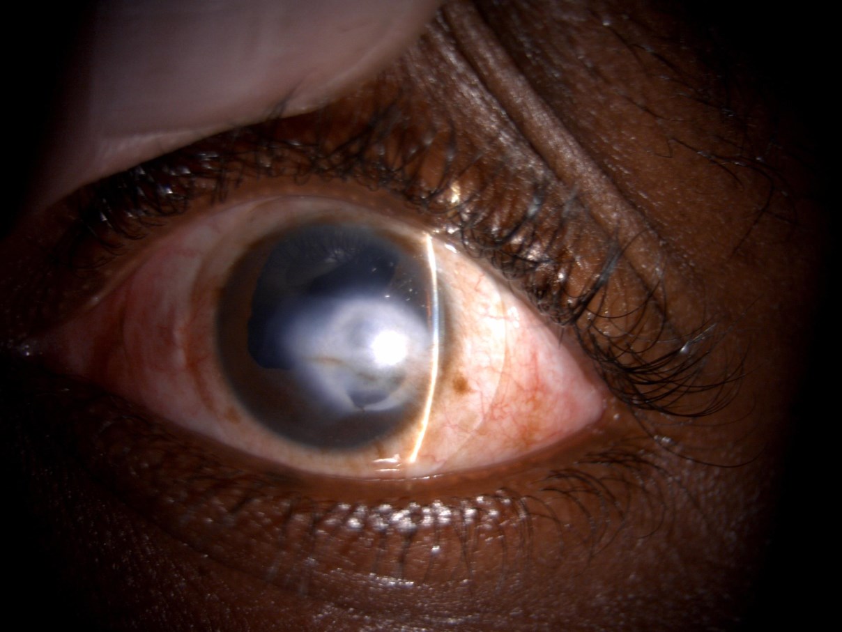
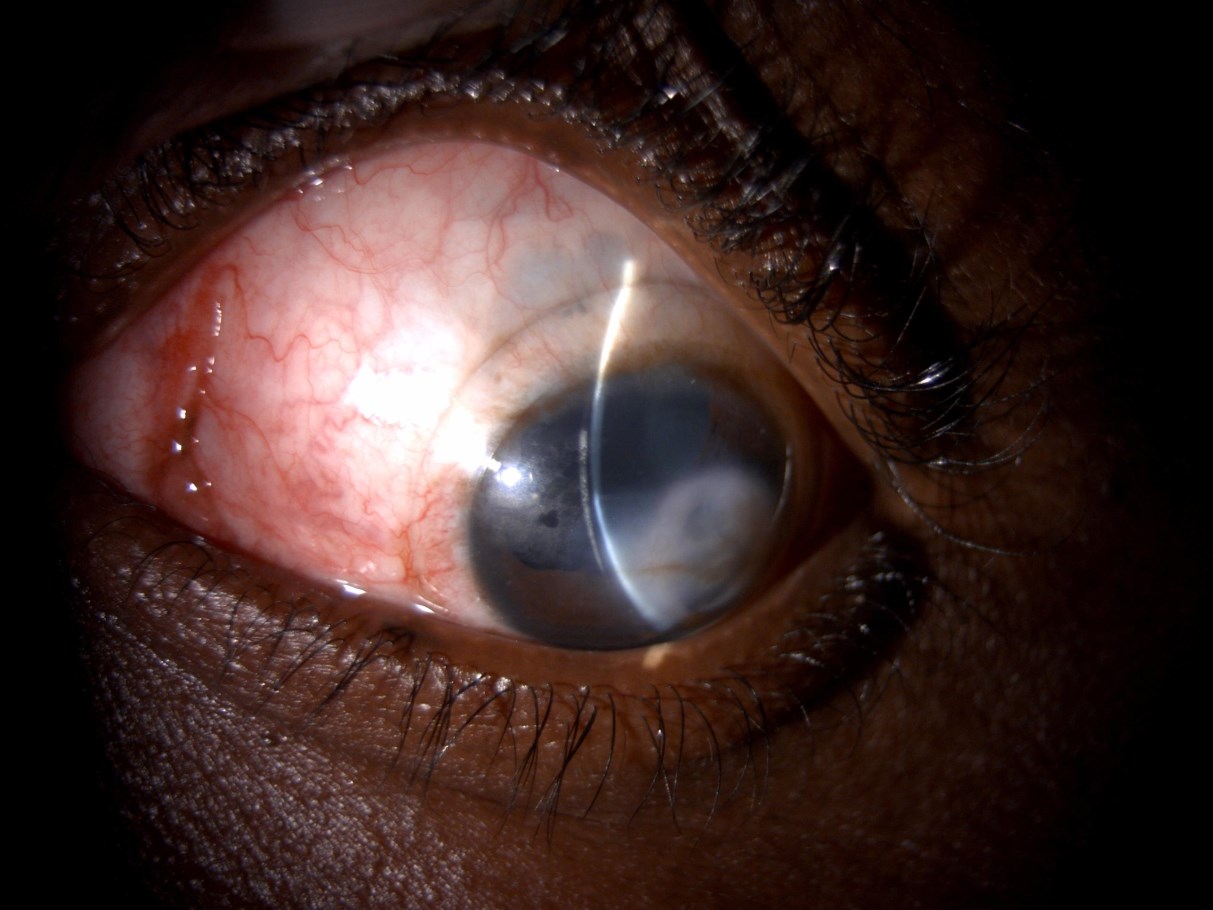
Thabo (pseudonym) is a 33 year old indigent African male with complaints of poor vision in both eyes since birth. At the initial visit on the 1st July 2013 he reported that he has never been able to see well and although the exact details are unclear he was told that he had an “eye infection” when he was born. He has no known history of other ophthalmic problems and is in good health. On examination he had severe scarring of both corneas (Figure 1). The right eye has a completely scarred vascularised cornea (generalised leukoma or wall eye) with no visualisation of any anterior chamber structures possible (Figure 3). Vision was light perception. I do not have access to B-scan ultrasound and could therefore not examine the intraocular structures and posterior segment. The left eye has severe vascularised scarring involving 80 % of the cornea (Figure 2 and 4). The central area has previously perforated and resolved with leukoma adherens and distortion of the iris (corectopia). The anterior chamber is well formed and there was no active inflammation. The intraocular pressure was 16 mmHg and both the lens and vitreous was clear. Mydriatic examination of the retina was not possible but it appears normal with indirect ophthalmoscopy. The disc is tilted with a cup to disc ratio of 0.4. Refraction was -12.00/-4.00×110 and vision with correction was 20/200. He fixates through the paracentral cornea which makes it difficult to establish an accurate refraction. The most likely cause of the corneal scarring is neonatal conjunctivitis/keratitis and corneal perforation due to Neisseria gonorrhoeae. It does not look like a congenital anterior segment dysgenesis. In terms of management a few options exist, including contact lenses for the management of complex corneal pathology and corneal transplant surgery. The option for a corneal transplant should only be considered as an absolutely last option due to the extensive degree of vascularisation which will significantly raise the chance of corneal rejection and graft failure. Common post-operative complications of penetrating keratoplasty include wound leaks, flat anterior chamber with increased IOP, endophthalmitis, persistent epithelial defects, primary graft failure, problems related to sutures, post-operative astigmatism, corneal ulcers, recurrence of disease, and graft rejection 1. The prevalence of endothelial graft rejection is around 21% 1. Symptoms include pain, photophobia, redness, and decreased vision.
Contact lens fitting and examination
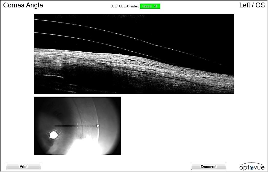
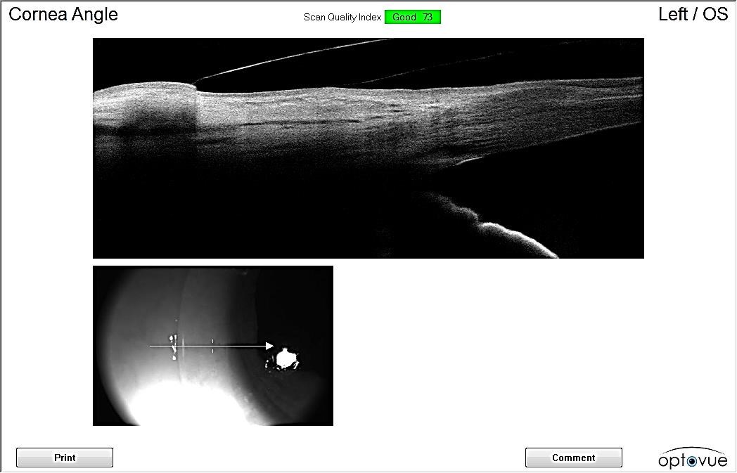
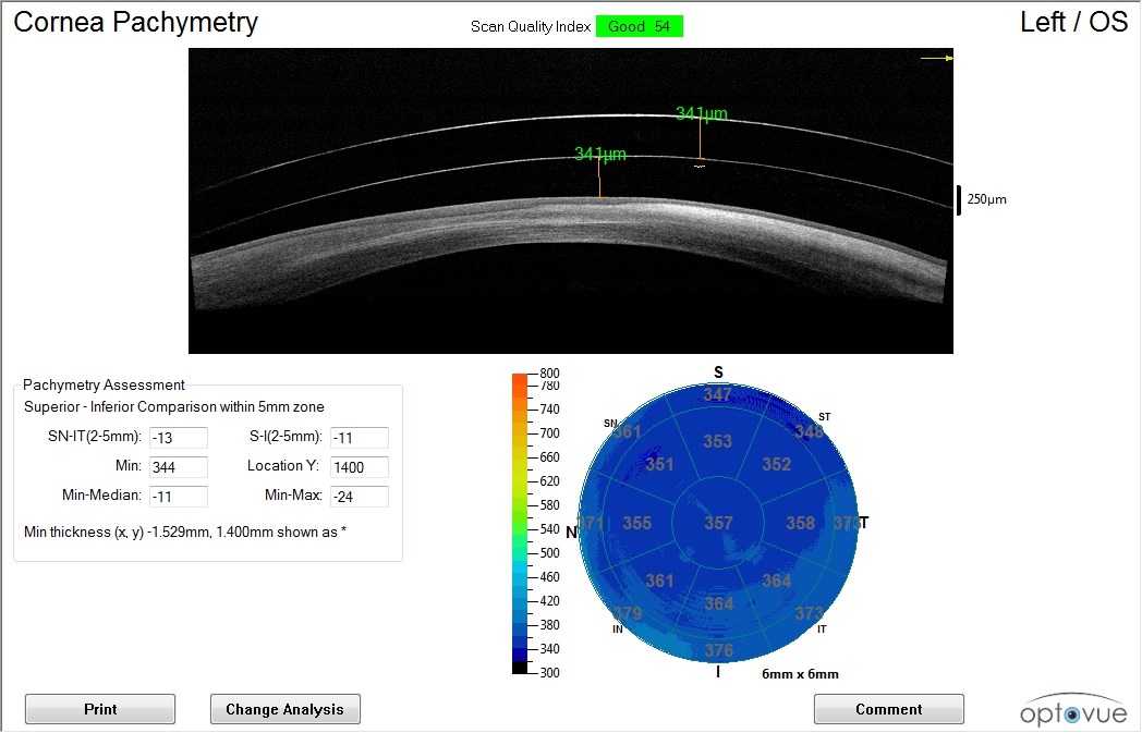
Several types of contact lenses were considered for Thabo. It was difficult to accurately analyse his cornea with the Pentacam due to his unstable fixation and degree of corneal scarring. A mini-scleral trial lens was selected and the fit was evaluated with the slit lamp and fluorescein. This process was repeated until a satisfactory fit was achieved. The final lens parameters are listed in figure 10 and the fluorescein pattern is shown in figures 1 and 2. Figures 4 to 9 show the fit of the lens as well as the apical clearance (estimated at 340 microns), limbal clearance (estimated at 215 microns temporally and 230 microns nasally) and scleral fit with anterior segment OCT. The lens did not bear on the cornea and cleared the limbus completely resting on the sclera. No blanching of the conjunctival vasculature surrounding the limbus was evident indicating a good scleral fitting relationship in all meridians. The final lens power was -1.50 and visual acuity 20/200. +3.50D reading spectacles as well as a 3.5x hand magnifier helped Thabo with his reading activities. He was instructed on different lens insertion and removal techniques and felt most comfortable using a DMV plunger to remove and insert the lenses. The lens bowl was filled with unpreserved saline (Alcon Polyrinse®) before insertion. Hydrogen peroxide was recommended as a disinfection system (AO Sept) after cleaning and rinsing the lenses with a surfactant (Crystal Cleaner– alcohol based surfactant) and saline respectively. Alcohol based surfactant cleaners are very effective in removing surface lipid and protein deposits with manual rubbing. In combination with hydrogen peroxide sterilization this cleaning regime is very effective for all types of contact lenses. Hydrogen peroxide was specifically selected for this patient due to his remote rural location and the significant risk of Acanthamoeba infection due to unsanitary living conditions and poor drinking water quality which makes the use of multipurpose solutions extremely risky. A two-step 3% hydrogen peroxide solution is 99.9% cysticidal providing contact times of at least four hours are employed 4. Hydrogen peroxide also provides excellent antimicrobial and antifungal efficacy. It works by penetrating the lens material and cleans by expanding the lens matrix (soft lenses) and oxidizing microbes. Because of its hypotonic nature and pH of 4.00, H2O2 is also able to break protein and lipid bonds, remove trapped debris and penetrate bacterial biofilms 5. H2O2 is highly effective against all microorganisms when used in a 3% concentration but is non-selective in its activity.
Although instructed to visit my office regularly for after care Thabo did not manage to see me until the 17th October 2015 (two years after the initial fitting) only because he needed a new lens. He lives in a rural community in the Orange Free State, about 400 km from my office. He has no fixed income and no transport. He relies on a state subsidy and transport arranged by the local clinic for his visits to my office. At the visit on the 17th October 2015 his examination revealed that the mini-scleral lens did not cause any adverse effects and his vision remained at 20/200. Retinal examination was as before and his IOP remained at 16mmHg. Corneal neovascularization have not increased and more importantly Thabo was extremely happy with the visual outcome achieved with the semi-scleral lens.
Differential diagnosis
Other than traumatic injury (chemical, surgical, penetrating) which leads to scarring, any disease which interferes with corneal clarity can lead to a corneal opacity 1,2,6.
“STUMPED classification”
- S – Sclerocornea
- T – Tears in Descemet’s membrane
- Congenital Glaucoma
- Birth Trauma
- U – Ulcers
- Herpes virus
- Bacterial
- Neurotrophic
- M – Metabolism (rarely present at birth)
- Mucopolysaccharidoses
- Mucolipidoses
- Tyrosinosis
- P – Posterior corneal defects
- Peter’s anomaly
- Posterior keratoconus
- Staphyloma
- E – Endothelial dystrophy
- Congenital hereditary endothelial dystrophy
- Congenital hereditary stromal dystrophy
- D – Dermoid
Discussion
Van Der Worp, 2015 classifies a mini-scleral lens as having a diameter of 12.5 to 15.00 mm sharing bearing on the cornea and sclera 7. This classification is supported by the Scleral Lens Education Society Nomenclature which defines a mini-scleral lens as a lens that rests entirely on the sclera and is less than 6 mm larger than the horizontal visible iris diameter 8. In this case a rigid gas-permeable mini-scleral lens was used to correct Thabo’s refractive error and corneal irregularity. Oxygen delivery to the cornea is impacted by the scleral lens and the tear lens thickness, especially if the scleral lens was thicker than 350 microns and the tear layer thickness exceeds 200 microns 9. In order to minimize hypoxia induced swelling with scleral lenses, lenses with high Dk values (>150), maximal centre thickness of <250 microns or less, and tear lens thickness not exceeding 200 microns should be used 9. This theoretical model has been validated clinically, large scleral lens wear results in 2-3% corneal oedema at the end of the wearing period if the lens and tear layer is thicker than 300 and 200 microns respectively 7. It has been suggested that this level of oedema is not clinically significant, being very similar to the oedema present upon awakening 10. Compan et al, 2014 also suggested that scleral lens materials should have at least a Dk of 125 with a thickness of 200 microns and a tear film thickness of 150 microns or less to meet oxygen tension of 55mmHg – which is considered the minimum critical barrier to avoid clinically significant hypoxia 11. More recently Jaynes et al., 2015 suggested that corneal oxygen consumption would be higher after a patient has been wearing thick rigid gas-permeable lenses with higher corneal clearances 12. Thinner tear lens and lens thickness seem to have a positive effect on the oxygen transmissibility of the system ensuring long term corneal health. However, care should be taken to ensure that the corneal clearance (vault) is maintained during lens wear. It is known that clearance decreases naturally due to “sinking” of the lens during wear into the conjunctiva and this should be kept in mind when fitting the scleral lenses 7. Rigid gas-permeable contact lenses are hydrophobic which causes them to be more prone to lipid than protein deposition on the lens surface. The bound lipids reduces the hydrophobicity of the lens surface allowing protein to bind and the bound deposited hydrophilic protein then alters subsequent binding of both protein and lipid making cleaning of high gas permeable lenses difficult 13. Another factor to consider is contact lens flexure. Lens flexure is directly affected by lens thickness and gas-permeability, thinner high gas-permeable lenses are prone to more flexure 14. Lenses that flex on the eye can affect the fitting characteristics as well as optical quality and therefore the vision negatively. Flexure can be confirmed by manual or automated keratometry or corneal topography over the lenses, any toricity detected will provide an index of lens flexure. A balance between the corneas oxygen requirements, lens oxygen transmissibility, lens thickness and lens wetting angle is needed to ensure optimal performance of a rigid gas-permeable semi scleral lens on the eye. Thabo’s cornea is severely vascularised and therefore great care needs to be taken to ensure the long term health and viability of this tissue. This means regular follow-up visits to carefully examine the cornea for signs of increased vascularisation and other signs of corneal problems related to semi-scleral contact lens wear.
Conclusion
Although Thabo only manages 20/200 acuity with the lens it makes a significant difference in his life. He now manages to do most of his house work and cooking on his own and can at least travel unassisted. Apparently he is a gospel singer and regularly travels throughout Africa to perform. I dispensed Thabo’s first lens in July 2013 and he has been using the lens without problems since that visit. Unfortunately he was mugged while visiting Angola recently and lost all his possessions which included a toiletry bag with his lens. This necessitated a visit to my office which would probably not have happened otherwise. After seeing him again a new lens was supplied and he is doing well. I have written to the rural medical clinic that assists him with transport instructing them that follow-up visits have been scheduled on a three monthly basis which he needs to keep. All work, lenses and solutions were supplied on a pro bono basis.
Figures
| Right | Radius | Diameter |
|---|---|---|
| Sag 4.3776 @15.60 mm | 7.84 | 8.60 |
| CT 0.25mm | 8.04 | 12.00 |
| 8.70 | 14.00 | |
| 13.00 | 15.00 | |
| 14.60 | 16.00 | |
| -1.50 | Boston XO2 Dk 141 |
References
- Yanoff M, Duker J. Yanoff: Ophthalmology. Elsevier Science Health Science Division; 1998.
- Kanski JJ, Bowling B. Clinical Ophthalmology: A Systematic Approach. Elsevier Health Sciences UK; 2011.
- Rours G, Hop W, Htun Y, et al. Carriage of chlamydia trachomatis during presgnancy: Consequences for mother and infant. The South AfricanJournal of Epidemiology and Infection. 2006;21(1):20-25.
- Hughes R, Kilvington S. Comparison of Hydrogen Peroxide Contact Lens Disinfection Systems and Solutions against Acanthamoeba polyphaga. Antimicrobial Agents and Chemotherapy. 2001;45(7):2038-2043.
- Gromaki SJ. Hydrogen peroxide disinfection. Contact lens Spectrum. 2006;21(12):19.
- Arffa RC, Grayson M. Grayson’s Diseases of the Cornea. Mosby; 1997.
- Van der Worp E. A Guide to Scleral lens fitting (2nd edition). 2015.
- Baldwin B, Jedlicka J. SCLERAL LENS NOMENCLATURE. 2013; https://www.sclerallens.org/sites/default/files/files/SLS_Nomenclature_LtrHead06_26_2013.pdf. Accessed January 2016.
- Michaud L, van der Worp E, Brazeau D, Warde R, Giasson CJ. Predicting estimates of oxygen transmissibility for scleral lenses. Contact lens & anterior eye : the journal of the British Contact Lens Association. 2012;35(6):266-271.
- Caroline PJ, McMahon J, Lotoczky J, Rosen C, Michaud L. Scleral lenses: new opportunities, new challenges. Global speciality lens symposium; January 2015, 2015; Las Vegas.
- Compan V, Oliveira C, Aguilella-Arzo M, Molla S, Peixoto-de-Matos SC, Gonzalez-Meijome JM. Oxygen diffusion and edema with modern scleral rigid gas permeable contact lenses. Invest Ophthalmol Vis Sci. 2014;55(10):6421-6429.
- Jaynes J, Weissman BA, Edrington T. Predicting scleral GP lens entrapped tear layer oxygen tensions. Contact lens & anterior eye : the journal of the British Contact Lens Association. 2015;38(5):392.
- Bontempo AR, Rapp J. Protein-lipid interaction on the surface of a rigid gas-permeable contact lens in vitro. Current eye research. 1997;16(12):1258-1262.
- STEVENSON RWW. Young’s Modulus Measurements of Gas Permeable Contact Lens Materials. Optometry & Vision Science. 1991;68(2):142-145.