
Dirk J. Booysen Dip Optom FOA(SA), MCOptom(UK), TMOD(USA),CAS(USA)
Part 1
The role of nutrition in eye care, especially in the treatment of AMD, has been a topic of discussion in the professional as well as the lay press of recent.
Although a plethora of research studies report strong evidence that antioxidant vitamins, caretenoids and herbs can significantly help lower the risk and slow the progression of wet and dry AMD, cataract formation and glaucoma in some patients, just as many studies dispute these findings.
The conflicting evidence has neither dampened patient curiosity about alternative medicine for eye health, nor hindered sales of nutritional products claiming to reduce the risk of ocular disease.
Sales of nutritional products in the USA top $ 18.7 billion dollars annually.
Furthermore, the level of understanding on matters relating to nutrition among ECP’s varies considerably as do opinions regarding the benefit if any to their patients suffering from devastating degenerative disease like AMD.
I am not a biochemist therefore some errors in interpretation of the information available on the subject are inevitable but not intentional. Further reading is therefore encouraged. However, this series of articles will humbly attempt to clarify a very complex and often extremely confusing subject.
The first article in the series which follows here will discuss the significance of Oxidative stress in the eye, and the second article will deal with the Nutrients, Nutrient derivatives and Herbs relevant to ocular health. The final article will deal with current research into Nutrition-related eye health, General Rules to supplement formulations, Contraindications and Adverse reactions to supplements as well as common drug interactions.
Oxidative stress and the eye
The process of oxidation is essential if a cell is to provide energy for vital cell functions. During this process 95 – 98% of the oxygen consumed is reduced to water, but the remaining 2 – 5% is converted to unstable and damaging ROI (reactive oxygen intermediaries).
At physiologic levels, ROI’s function as signaling and regulating molecules, but at pathologic levels they are highly deleterious and act as cytotoxic oxidants. Prolonged exposure to ROI results in DNA mutation, tissue injury and disease.
However, the body has an inherent defensive system, consisting of antioxidants and antioxidant enzymes, which act synergistically in scavenging the ROI and thus protecting the underlying tissues.
Oxidative stresses occur when the level of oxidants in the system exceeds the detoxifying capacity of its antioxidants; leading to oxidative damage to macro molecules with consequential injury to cells and tissue.
Chemical reactions which involve Oxidation and Reduction of molecules occur in every cell. Oxidation referring to the removal of electrons and Reduction to the gain of electrons.
The majority of ROI’s are formed during energy generation from mitochondria, or during detoxifying reactions. However, pollution, asbestos, fungal or viral infections, cigarette smoking, excess alcohol consumption, ultraviolet irradiation, inflammation and ageing are all known to be associated with increased production of ROI’s.
ROI’s can be classified according to their reactivity toward biological targets, their site of production, their chemical nature, or their free radical or non-radical subgroups.
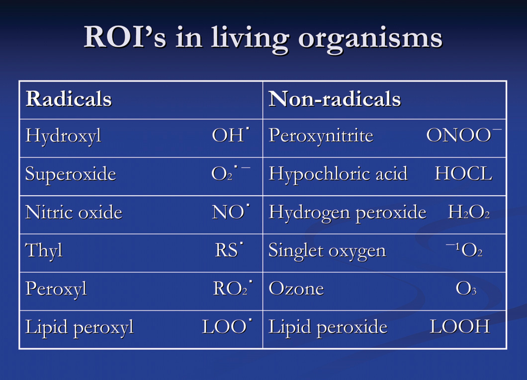
Free radicals are molecules that have a single unpaired electron in their outer orbit. They are very unstable (catalytic molecules) and react with nearby molecules. In order to achieve a stable state, free radicals extract electrons from other molecules, which are then rendered unstable, resulting in cytotoxic chain reactions.
Cell membranes are composed of abundant lipid. Oxygen-derived free radicals react with lipid ( EFA’s) in cell membranes. Unsaturated fatty acids are highly susceptible to oxidation and free radical damage, because their conjugated double bonds are convenient sources of hydrogen atoms, which contain one electron.
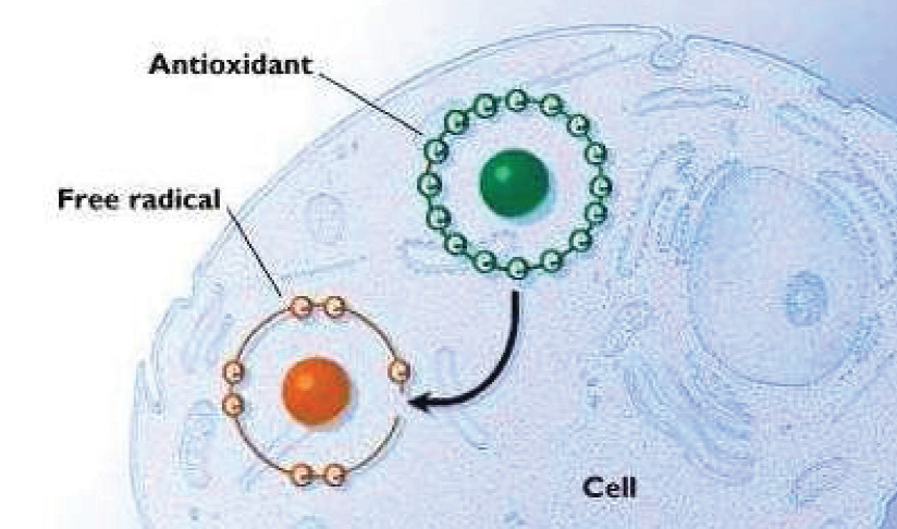
Certain free radicals are used by cells in the body for protection against bacteria.
Non-radical ROI’s contain their full complement of electrons, but in an unstable state. The most important non-radical ROI’S are Hydrogen Peroxide and Singlet Oxygen.
Every component of the eye is vulnerable to damage from ROI, however the retina is particularly susceptible. The reasons for this include high concentrations of polyunsaturated fatty acid (PUFA) in the outer segments of the photoreceptors which are readily oxidized, constant exposure to visible blue light (cornea filters most ultra violet), high consumption of oxygen by the mitochondria in the inner segments of the photoreceptors forming and leaking ROI, increased partial pressure of Oxygen, an abundance of photosensitisers (lipofuscin) in the neurosensory retina and the RPE, and the process of phacocytosis of the photoreceptor outer segments by the RPE which is known to generate Hydrogen Peroxide.
Therefore photoreceptors find themselves in a high risk pro-oxidative environment.
The antioxidant defense system consist of enzymatic (endogenous) and exogenous components, cellular compartmentalization and repair.
Antioxidants are substances that significantly delay or prevent oxidization of a substrate.
Endogenous Antioxidants
Endogenous Antioxidants include the enzymes, glutathione peroxidase (GSH), superoxide dismutases (SOD) and catalase. Glutathione peroxidase is a universal antioxidant, abundant in the cytoplasm, nuclei and mitochondria of cells. It is dependent on selenium (Se) as a cofactor.
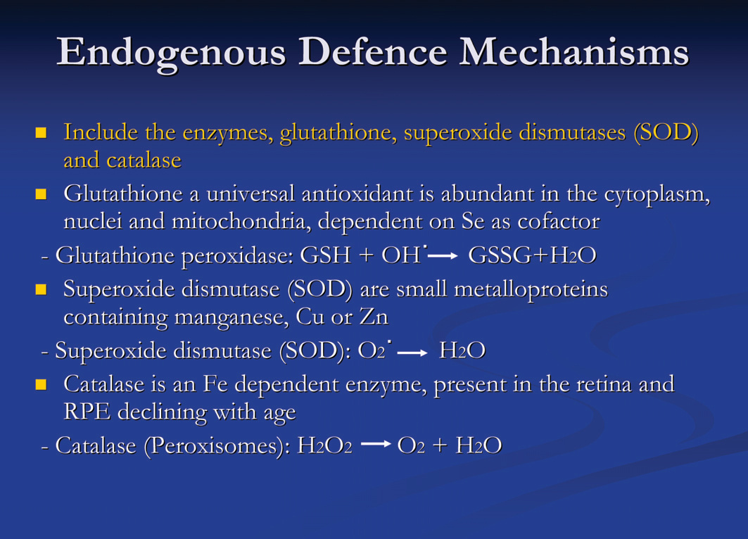
Superoxide dismutases are small metalloproteins, some containing maganese, others Copper or Zinc. SOD catalyses the quenching of superoxide to produce water.
Catalase is an iron-dependent enzyme that scavenges H2O2 either catalytically or perioxidatively, producing oxygen and water. It is present in the retina and RPE, declining with age.
Exogenous Antioxidants
Include vitamins A, C, E, carotenoids, bioflavonoid, selenium and zinc.
Vitamin A protects the photoreceptor membranes against oxidative damage by breaking the chain reaction during lipid per-oxidation. It is also involved in the repair of cells that have been injured by oxidation.
Vitamin C (ascorbate) is a major water-soluble antioxidant. Water solubility makes it extremely effective in ocular tissue which has a higher concentration of Vitamin C than plasma.
Vitamin E (tocopherol) act synergistically with carotenoids in scavenging free radicals.
Macular pigment (yellow spot in the centre of the retina) is composed of two hydroxycaretonoids, lutein and zeazanthin. MP is entirely of dietary origin and is found in high concentrations in green leafy vegetables, fruits and egg yolk.
The function of MP is to filter harmful ultraviolet light, protecting the retina from photochemical damage and to act as free radical scavenger. In addition it also reduces chromatic aberration.
Bioflavonoid (vitamin P) is large polyphenolic molecules which are derived from the peel and covering of teas, berries, grapes and bark. Bioflavonoids are entirely dietary dependent and the most common form is Quecetrin. They exhibit a myriad of properties, such as anti-inflammatory, antibacterial, and antioxidant activity. Sources include red wine, green tea and English Blueberry, with green tea producing the most potent antioxidant known to humans. Bioflavanoids also protect Vitamin C from oxidation and aid in its absorption, allowing the body to reap more benefit from Vitamin C.
Normally the production of ROI is met by an ample supply of the detoxifying enzyme SOD (superoxide dismutases) whereas catalase and glutathione reductase are readily available to cope with hydrogen peroxide.
These enzymes, in concert with antioxidant proteins and small molecular reductants, form an effective antioxidant defense within the photoreceptor. When overwhelmed, lipid peroxidation within the outer segment results. Protein makes up the remaining 50% of the lipid bilayer of the rod outer segment. Fragmentation, cross-linking and agepregation of proteins as well as enhanced vulnerability to proteolysis, can result from oxidation of their amino acids. The oxidised basis of DNA, arising from interactions with ROI is believed to contribute significantly to ageing and age-related disorders involving many organ systems, including the eye.
Oxidative stress and age related macular degeneration
In western countries, AMD is the leading cause of blindness in people over 65. In the USA as many as 13 million people have evidence of AMD, impairing about 1.2 million people. 30% of people over 75 have AMD, 23% of the remainder will develop AMD in the next 5 years of life.
There is currently no cure for “Dry” or atrophic AMD, the form characterized by hard or soft drusen (deposits or cellular debris), changes in retinal pigment epithelium, or atrophy of photoreceptors and the retinal pigment epithelium.
The atrophic form of AMD accounts for approximately 90% of all cases of AMD. Less than 20% of all “Wet” AMD cases are candidates for photocoagulation. Recently major advances in the treatment of “Wet” AMD in the form intravitreal injections of antiangiogenic drugs Lucentis or Avastin, and photodynamic laser therapy have been made which hold promise for future treatment.
The exact cause of AMD still remains elusive; however, oxidative stress has been increasingly linked to its aetiopathogenesis.
Evidence of oxidative stress can be seen in the RPE and neurosensory retina with increasing age, and this damage is most prominent in the region of the retina where AMD changes are seen. The RPE metabolically maintain and support the photoreceptors, therefore RPE dysfunction contributes to the pathogenesis of AMD. Bruch’s membranes hydraulic conductivity or waste product pump is reduced leading to RPE detachments. Debris accumulates in Bruch’s membrane, altering its composition and permeability further. The RPE’s ability to phagocytose rod outer segments is impaired, resulting in an accumulation of lipofuscin with eventual geographic atrophy.
Lipofuscin is a generic name given to a heterogeneous group of complex, auto fluorescent lipid and protein aggregates present in neural and non-neural tissue, resulting from oxidatively damaged cells.
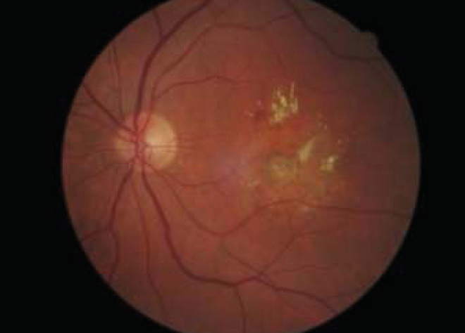
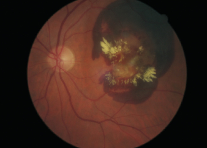
Lipofuscins have three primary defining characteristics;
- They consist of intracellular secondary lysosomes
- They have yellow auto fluorescence when excited by UV or blue light
- They accumulate during normal ageing
High concentrations of Lipofuscin accumulate in the vicinity of the RPE and Bruch’s membrane near the end of the lifespan, which can not be removed, resulting in progressive cellular dysfunction. Normal autographic processes are interfered with, resulting in accumulation of damaged cellular constituents. Lipofuscin also sensitizes lysosomes and neurons to oxidative stress by binding with metals such as Cu and Fe. Lipofuscin is a photo inducible generator of ROI. Exposure to blue light produces superoxide ions, singlet oxygen, hydrogen peroxide and lipid peroxides. While there is increasing indirect evidence that the accumulation of lipofuscin is involved in the development of AMD, the fundamental question on whether or how it results in impairment of cellular function remains.
In response to this increased oxidative and metabolic stress, the RPE produces VEGF and fibroblast growth factor, which may stimulate new blood vessel growth and CNVM formation.
Oxidative stress and cataract
Exogenous UV light and reduced endogenous antioxidant enzyme activity can generate ROI’s in the lens rendering it vulnerable to protein modification, lipid peroxidation and DNA fragmentation. With age the protection mechanism against oxidation in the lens, glutathione, superoxide dismutases and catalase slowly deteriorates and becomes ineffective leading to cataract formation.
Oxidative stress and glaucoma
This theory postulates that raised IOP in POAG is attributable to malfunction of the trabecular meshwork (TM) and Schlemm’s canal outflow system. The TM is believed to be exposed to chronic oxidative stress because of the presence of ROI in the aqueous humour. The generation of ROI is mainly due to mechanical stress and intracellular metabolism.
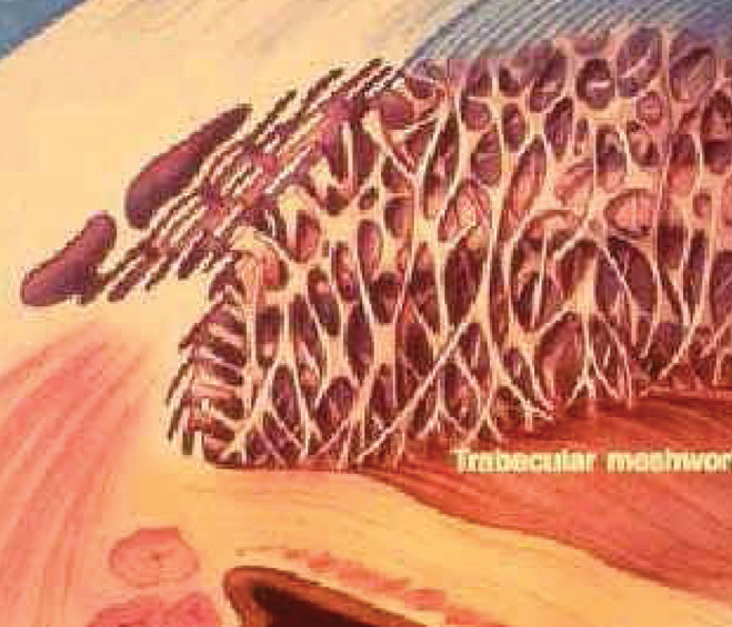
The TM posses several protective mechanisms to deal with oxidative stress including high levels of superoxide dismutases, glutathione, catalase, ascorbate (vitamin C), tyrosine, as well as proteasome which eliminate proteins altered by ROI.
In POAG the levels of glutathione, ascorbate and tyrosine are significantly reduced.
Furthermore proteasome can be impaired by the following mechanisms.
- Saturation of the proteasome by the presence of excessive number of altered proteins.
- Alteration of proteasome by direct oxidation leading to impaired function.
The capacity of the TM to protect itself from oxidative damage has been proposed to underline the pathophysiology of POA
- 98% of oxygen is converted to water and 2% is converted to harmful ROI during mitochondrial metabolism
- Other sources of ROI include, pollution, asbestos, infection, inflammation, smoking, alcohol, ageing and ultra violet radiation
- High levels of ROI’s are cytotoxic, causing DNA mutation, tissue injury and disease
- Most common classification of ROI’s are free radicals or non-free radicals
- Free radical molecules have single unpaired electrons in outer orbit and react readily with PUFA’s in cell membranes – lipid peroxidation
- Non-radicals have their full complement of electrons but are unstable, damaging molecules when converting to another state. Most important non-radicals are; Hydrogen peroxide and Singlet oxygen
- Defense mechanisms against oxidative damage include endogenous enzymes, SOD, GSH and catalase and exogenous antioxidant vitamins A, C, E, the carotenoids, bioflavonoids, minerals and trace elements Zn, Se, Fe, Mn and Cu
- The retina is highly susceptible to ROI damage due to high concentrations of PUFA’s in the photoreceptor outer segments, constant exposure to visible blue light, high consumption of oxygen, high partial pressure of oxygen, abundance of lipofuscin, and phacocytosis of photoreceptor outer segments generating hydrogen peroxide
- It has been postulated that oxidative stress, blue light exposure, and the lack of endogenous and exogenous defense mechanisms play a role in the pathogenesis of AMD, Cataract formation, POAG and other degenerative eye disease
Figure 7 Summary, Oxidative Stress
References:
- Nutritional and environmental influences on the eye, edited by Allen Taylor, CRC press
- Nutrition and the eye, a practical approach, edited by Frank Eperjesi and Stephen Beatty, Butterworth Heineman Elsevier
- Biochemistry of the eye, 2nd edition, David R. Whikehart, Butterworth Heineman
- Prescription for nutritional healing, third edition, Phyllis A. Balch, James A. Balch, Avery
- Biochemistry, International edition, Mary K. Campbell, Saunders colledge publishing
- The Merck Manual, 16th edition, MSD
- Concise Physiology, HS Meij, DH van Papendorp, Department of physiology UOFS
- The seeds of good health, Montgomery Vickers, Review of Optometry, Online
- Vitamins and age related macular degeneration, Shantan Reddy, Howard F. Fine, July/August 2006, Eyecare Africa
- What we know about AMD and Nutrition, Stephen G. Pratt, Review of Ophthalmology, Online
- The Nutrition Connection: The news on diet and eye diseases, Jefferey S. Eisenberg, Review of optometry, Online
Part 2: Nutrient, nutrient derivatives and herbs relevant to ocular health
Eating is far more complicated than simply warding off hunger or having a pleasurable experience. It involves aspects of our psychological make-up, social group, mood and many external factors relating to the availability of food. Eating also does more than just keep us alive. When insufficient food or specific nutrients are supplied, some physiological adaptation may occur to minimize the consequences. Eventually, however a deficiency state will arise.
At the beginning of the 20th century the science of nutrition was directed at discovering the essential nutrients, studying the effects of insufficient intakes and determining the quantities needed to prevent deficiency states.
Since then it has been gradually realized that good nutrition is not simply a matter of providing enough of all nutrients. We now realize that diets in affluent developed countries, although apparently containing all the necessary nutrients, are probably contributing too many of the diseases afflicting these populations. Much research is focused on finding which nutrients are linked to which diseases, in an effort to promote a change in the dietary intake and hence an improvement in health.
In this second article on nutrition I will review the macro and micronutrients relevant to ocular health, as well as some herbs commonly used in OTC formulations.
Macronutrients
The macronutrients include proteins, carbohydrates and the lipids. The lipids can be further subdivided into fatty acids, triglycerides and phospholipids.
Proteins:
Proteins are polymers of amino acids linked by peptide bonds. These polymers are arbitrarily called peptides when their molecular weight is below 10 000 and are referred to as proteins when their molecular weight exceeds 10 000. Proteins serve many vital roles in cells and tissues of biological constituents from viruses to man. Such roles include:
- Mechanical support
- Control of growth and differentiation
- Catalysis
- Transport and storage
- Motion
- Nerve propagation
- Immune protection
In the eye, proteins are known to:
- Support the structure and clarity of the cornea
- To participate in the variable light refraction of the lens
- To initiate transduction of light into electrical signaling
- To generate intraocular pressure
- To lyse bacteria in the pre-corneal tear film
- And other functions to indirectly sustain vision
Although there are a huge number of proteins in the eye, some deserve special attention. They include retinal rhodopsin, lens crystallins, cone pigment proteins, mucus glycoproteins, collagen and enzymes. (Figure 8)
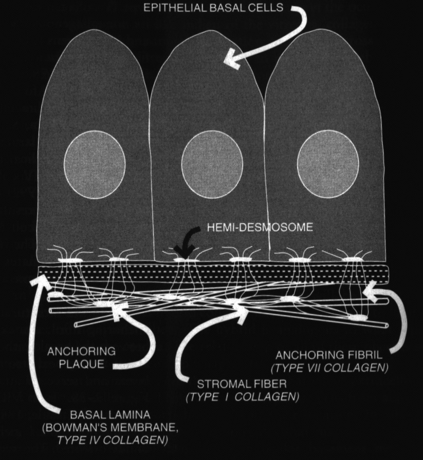
Crystallins are soluble lens proteins whose normal function is supportive in the maintenance of elongated lens fiber cells. These proteins are considered to be involved in the manifestation of senile cortical cataracts by oxidation of the disulfide bonds (process incompletely understood). They have also been implicated in the formation of nuclear cataracts. Rhodopsin and cone pigment proteins act as the initial participants in phototransduction. They are membrane proteins found on the discs of the rods and cones. Vitamin A (retinal) release triggers the cascade of phototransduction. Mucus glycoproteins known as mucins are found in the tear film and act to stabilize the tear film. Collagen composes the major type of ocular protein and is found in 80 to 90% of the bulk of the eye. It forms complex structures from basic tropocollagen units, which may form fibers, ground substances, or anchoring rods. It is also a constituent of the vitreous humour.
Enzymes are proteins that act as biological catalysts for a variety of cellular and extra-cellular reactions. In the eye, enzymes promote many of the same reactions found in other parts of the body. However, some enzymes have specialized activities related to ocular function and repair. For example, lysozyme acts in the precorneal tear film to destroy gram-positive bacteria by hydrolysis of their peptidoglycan coats. The sodium, potassium activated enzyme ATPase acts in the corneal endothelium to maintain corneal hydration, and in the cilliary body it generates intraocular pressure.
Sources of protein is divided into complete proteins containing ample amounts of all the essential amino acids, found in meat, fish, poultry, cheese, eggs, and milk. Incomplete proteins contain only some of the essential amino acids and are found in grains, legumes, and leafy green vegetables. Mutual supplementation enables combinations of partial-protein foods to make complementary protein, which supply all the essential proteins without the high fat content of animal protein. Examples are combinations of brown rice and beans and brown rice and nuts.
A complete discussion of the proteins is beyond the scope of this article, further reading is encouraged.
Carbohydrates:
Carbohydrates are divided into two groups, simple and complex carbohydrates. Simple carbohydrates or simple sugars, include fructose (fruit sugar), sucrose (table sugar), and lactose (milk sugar), as well as several other sugars. Complex carbohydrates are also made up of sugars, but the sugar molecules are strung together to form longer, more complex chains. Complex carbohydrates include fibre and starches, which include vegetables, whole grains, peas, and beans. Carbohydrates are the main source of blood glucose, which is the main fuel for all the body’s cells, and the only source of energy for the brain and red blood cells. Except for fibre, which cannot be digested, both simple and complex carbohydrates are converted into glucose. Glucose is either used directly to provide energy for the body or stored in the liver for future use. If more calories are consumed than a person uses, carbohydrates can be stored in the body as fat.
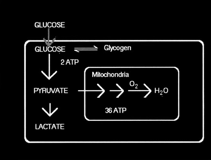
Ocular tissues use carbohydrates in monosaccharide form as sources of cellular ATP. The glycolytic reactions that are involved can occur in the presence or and absence of oxygen although some oxygen are always required for cell survival. Ocular cells use varying proportions of aerobic and anaerobic metabolism in glycolysis to achieve their particular energy demands. (Fig 9) Photoreceptors require the highest levels of ATP and lens fibre cells the least.
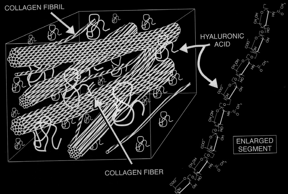
Structural carbohydrates (polysaccharides) form part of tissue structures in the extra cellular matrix. These derivatives are classified as glycosaminoglycans (GAG’s). Many GAG’s combine with core protein to form proteoglycans. In the eye these polymers are found in the vitreous, cornea, sclera, lens capsule, and blood vessels. (Figure 10.)
Lipids (Fats):
The terms lipid and fats have been used interchangeably to refer to groups of compounds that are water insoluble, but soluble in nonpolar solvents such as benzene, chloroform, alcohol and hexane. Strictly speaking, however, the term fat refers to lipid esters of fatty acids and glycerol. Lipids are hydrophobic and associate together in an aqueous or polar environment. It turns out however that lipids are also partially hydrophilic. This dual property of more dominant hydrophobicity and less dominant hydrophyllicity is called an amphipathic property. These characteristics make lipids very useful in cell membranes where both solubility properties are needed.
The major role of lipids in ocular tissue is as key components of membrane structure. Membrane lipids compromise the matrix that gives form and structure to membranes, and in which membrane proteins are imbedded. In the retina, the disc membranes of the outer segments of the photoreceptor cells play an important role in the vision process.
Lipids provide the maximum amount of energy (9 kcal/g) to the body, twice that of protein or carbohydrates. Lipids are emulsified by salts of bile acids and hydrolysed by lipases (enzymes) in the small intestine to fatty acids and glycerol. From the small intestine, hydrolysed lipids enter the blood stream, are transported to the liver where further metabolism takes place. Lipids facilitate intestinal absorption and transport of fat soluble vitamins A, D, E, and K.
Although not exhaustive, the most important lipid classes are; fatty acids, triacylglycerols (triglycerides), phospholipids, isoprenoids, esters, eicosanoids, steroids, and glycolipids.
Fatty acids.
There are three major categories of fatty acids, depending on the number of hydrogen atoms in their chemical structure.
- Saturated fatty acids. Primarily in animal products, including dairy items, such as whole milk, cream, cheese, and fatty meats like beef, veal, lamb, pork, and ham. Some vegetable products including coconut oil, palm kernel oil, and vegetable shortening are also high in saturates. Saturated fats are solid at room temperature, they are metabolised in the liver to cholesterol. High intake of saturated fatty acids can significantly raise the low density lipoprotein (LDL) level in the blood. Intake should not exceed 10% of total caloric intake.
- Polyunsaturated fatty acids. Found in the greatest abundance in corn, soybean, sunflower oil, and certain fish oils. Polyunsaturates can lower the total blood cholesterol; however, the high density lipoprotein (HDL) is also reduced. Intake should not exceed 10% of total caloric intake. Polyunsaturated fatty acids are also known as Vitamin F and although many fatty acids can by synthesised in the body, essential fatty acids (EFA’s) cannot and must be provided in the diet. Some of the important fatty acids in ocular tissue include arachidonic acid, linoleic acid, linolenic acid, and cervonic acid, which respectively play roles in the prostaglandin and leukotrine production, as well as visual transduction. EFA’s are classified into two families, Omega 3 and Omega 6 according to the number of carbon atoms, double bonds and the proximity of the first double bond to the methyl terminus of the acyl chain. For example, Linolenic acid = C18:3 Ω-3, has an 18 carbon chain with three double bonds, the first double bond at carbon 3.
Omega 3 essential fatty acid include alpha-linolenic and eicosapentaenoic acid. Ω-3 is found in fresh deepwater fish, fish oil, vegetable oils including canola, flaxseed and walnut oil. Omega 6 essential fatty acids include linoleic and gamma linoleic acid, found primarily in raw nuts, seeds, legumes and unsaturated vegetable oil, primrose oil and sesame oil.
Cervonic or docosahexaenoic acid (DHA), C22:6 Ω-3, is an essential fatty acid found in high concentrations in the brain, synaptosomes, sperm and highest concentration in the rod outer segments of the retina (ROS). It is synthesised from linolenic acid, primarily in the liver, then packaged into LDL’s for delivery to the retina. DHA is directly available from fish, fish and marine mammal oil and eggs, but not from flaxseed or vegetable oil which require an extra metabolic step. Once in the retina DHA is incorporated into phospholipids and the photoreceptor disc membranes. Here it associates with rhodopsin enabling its activation, hence facilitating visual transduction. DHA in the ROS is however highly unsaturated and therefore very susceptible to oxidation and ROI damage (see part one). Diets low in Omega 3 lead to impaired visual acuity and abnormal ERG which may not be reversible.
The following table summarizes the role of EFA’s in ocular health.
- Dietary intake of polyunsaturated EFA affects the overall amount of inflammatory activity in the body
- EFA’s are natural modulators of inflammatory processes via their metabolism to eicosanoids – Prostaglandin and Leukotrines
- Prostaglandins & leukotrines are involved in the control of inflammatory and immune responses
- This makes them useful in the treatment of dry eye, a chronic inflammatory condition
- Most importantly, linolenic acid, a precursor to DHA, is only available from the diet and plays a vital role in visual transduction
- Monounsaturated fatty acids. Are found mainly in vegetable and nut oils such as olive oil, peanut oil, and canola. They appear to lower LDL without affecting HDL in anyway. Intake should be kept at 10-15% of caloric intake.
- Triglycerides are lipids that represent a storage form of fatty acids. One molecule consists of three fatty acids bonded to a glycerol molecule using ester bonds. Most triglycerides are kept in fat cells and represent a large depot of stored energy, as well as a source of heat insulation. Triglycerides can be broken down to form acetyl CoA in order to obtain ATP. In the eye, however, triglycerides are used to maintain cell membranes and not so much as an energy store.
- Phospholipids, represent the most important lipid class for the formation and maintenance of cell membranes. The structure is similar to that of triglycerides, with both forms using glycerol as the frame on which esters are attached. In phospholipids a phosphate ester is used to bond the glycerol to the fatty acids. Cells design the phospholipids according to their functional needs with the hydrophobic fatty acid components determining the fluidity of the membranes. In the retina and photoreceptors the phospholipids contain high concentrations of highly unsaturated Cervonic acid which impart considerable fluidity to the cell membranes, vital to visual transduction.
- Isoprenoids. This lipid group represents a family of lipids metabolically built up from five-carbon units known as isoprene. Members of this class include.
- Cholesterol and its allied steroids such as the hormone cortisol
- Lipid soluble vitamins such as vitamin A, discussed later in this article
- Coenzyme Q
- Variety of essential oils in the plant world such as eucalyptus oil
- Cholesterol is an important lipid for a variety of other reasons besides its participation in membrane rigidity. It is a source of cholesterol esters, which are important components of the pre-corneal tear film. It is a synthetic precursor of a variety of steroid hormones that affect both ocular function and dysfunction. In ocular membranes where fluidity is important, such as the ROS, cholesterol is restricted to around 8% of the lipids in the disc membranes.
- Eicosanoids, are cyclic lipids derived from eicosanoic acids such as arachidonic acid. They include prostaglandins, thromboxanes, and leukotrines. They are short acting local hormones.
- Glycolipids, are important membrane components found in nervous, ocular and other tissues. Glycolipids are lipids that contain carbohydrates, such as galactose.
- Esters, are the bonds between a carboxylic acid (fatty acid) and either an alcohol or a hydroxyl group attached to a ring compound. Examples are the bonds formed between glycerol and fatty acids in the formation of triglycerides and phospholipids. Another type of lipid ester is represented by waxes. Waxes (esters of long-chain fatty acids and long-chain alcohols that are derived from fatty acids) are usually solid at room temperature and occur in nature as the shinny covering of plant leaves, bees wax, and the oily substances covering skin, hair, fur, and wool. In the eye, waxes are a major component of the lipid layer of the precorneal tear film and exist as liquids at the temperature of the tear film at around 35°C
Micronutrients
Like water, carbohydrates, protein, and fats, and the enzymes required to digest them, vitamins and minerals are essential to life. They are therefore considered nutrients, and are referred to as micronutrients simply because they are needed in relatively small amounts compared with the four macronutrients.
Vitamins protect cells and tissues from the detrimental effects of physical, chemical and microbial agents. The protective action is attributable to the maintenance and/ or augmentation of the body’s existing defense systems. Vitamins form an essential component of a balanced diet as they cannot be synthesized de novo in humans.
The body needs 13 different vitamins. Of these four are fat soluble (Vitamins A, D, E, and K), and nine are water soluble (The C and B group). Fat-soluble vitamins differ from water-soluble vitamins in several respects. Water-soluble vitamins are readily absorbed from the diet, and do not require transport proteins in the plasma. Fat soluble vitamins are stored in the liver and adipose tissue, and therefore can be mobilized during periods of deprivation. In contrast, deficiency states of water- soluble vitamins can occur after a relatively short period of deprivation (Scurvy)
Some vitamins, such as A, C and E, serve as antioxidants, and protect cells against free radical damage. Free radicals are ubiquitous in the human body, and disrupt the physiological functioning at a physiological level. The antioxidant vitamins act synergistically with antioxidant enzymes in scavenging free radicals, and their site of action can be intracellular or extracellular. (Part 1)
Vitamin A
Vitamin A earned its name from the fact that it was the first vitamin to be discovered. It is a generic term for a large number of related compounds, and can be categorized into two main groups: pre-formed vitamin A (retinoids) and pro vitamin A (carotenoids).
Retinoids:
Retinoids include retinol, retinal, retinoic acid and related compounds. Retinoids are found entirely in food sources of animal origin, and is the most reduced form of vitamin A satisfying all its known functions. Vitamin A is fat soluble and dietary fat must be consumed with sources of vitamin A to ensure absorption and bioavailability. Very low fat intake (< 15% of total energy) reduces the bioavailability of vitamin A. The richest sources of retinoids are beef and chicken liver, other sources include whole milk, butter and cheese.
Vitamin A is hydrolyzed in the intestinal mucosa, releasing retinol and free fatty acids. Retinol is esterified to palmitic acid, incorporated into chylomicrons and delivered to the blood. Chylomicron remnants are taken up by the liver resulting in storage of vitamin A within the lypocytes as retinyl palmitate. Transport to the other tissues including the outer retina, requires a binding protein (RBP – retinol binding protein). The RBP complex attaches to membrane sites at the RPE, RBP and albumin is cleaved from the retinol, which the passes into the RPE cell and eventually to the apical RPE cell membrane. It finally reaches the photoreceptor cells where retinol gives rise to visual pigment rhodopsin, and is used in the visual cycle.
Substances that affect vitamin A bioavailability include high doses of ferrous sulphate (iron supplements), tannic acid (black tea), aspirin, and nitrates from processed meats. Zinc deficiency interferes with vitamin A metabolism in several ways. Firstly, there is a decrease in the synthesis of RBP which reduces transport of retinol through the circulation. Secondly, there is decreased activity of the enzyme that releases retinol from its storage form (retinyl palmitate) in the liver. Thirdly, there is decreased conversion of retinol to retinal.
The principal forms of preformed vitamin A in supplements are retinyl palmitate and retinyl acetate.
Our Dermatology colleagues know that vitamin A is an essential factor in epithelial cell growth and cell differentiation. Retinoid deficiency enhances keratinisation of squamous epithelium and retinoid excess suppresses it. Retinoids can also change keratinized skin epithelium back into a form capable of secreting mucous.
Vitaminosis A results from malnutrition and patients present with conjunctival xerosis, Bitot’s spots and corneal softening. Symptoms include dry sandy feeling eyes and a marked decrease in night vision. These patients are treated with large doses of vitamin A (20 000 IU/day) and topical retinoic acid.
Carotenoids:
Carotenoids refer to a class of fat soluble colored pigments. They include β-carotene and related compounds, and are limited to sources of plant origin closely associated with chlorophyll and therefore play a critical part in photosynthesis. Yellow and orange vegetables and fruits are good sources. Although green vegetables also contain carotenoids they are masked by the presence of the green pigment chlorophyll. They are also found in non-photosynthetic microorganisms, where they protect against the detrimental effects of excess light and oxygen. A similar action exists at the macula in the human eye. Carotenoids cannot be synthesized de novo in the body, and are therefore entirely of dietary origin. Furthermore carotenes cannot be converted to xanthophylls, and vice versa. Of the approximately 600 carotenoids that occur in nature only 10% are pre cursors of vitamin A. Absorption of the carotenoids takes place in the duodenum and is dependent on the action of existing bile and pancreatic lipases. Passive transport into mucosal cells and blood circulation result in uptake by the liver and re-secretion on plasma lipoproteins (predominantly HDL).
Heating food influences the bioavailability of carotenoids by denaturing the carotenoproteins, and improving accessibility. However excess heating reduces availability, 60% of xanthophylls and 15% of carotenes are destroyed during the cooking process.
Carotenoid functions:
In humans carotenoids can serve several important functions. The most widely known is their provitamin A activity. The exact role of the two macular carotenoids or xanthophylls remains elusive. However, the functions of blue light filtration and antioxidant activity are particularly attractive in maintaining macular health.
The carotenoids can be subdivided into carotenes, and their derivatives xanthophylls.
Carotenes:
Carotenes are hydrocarbon carotenoids and include β-carotene, a-carotene, and lycopene. B-carotene, the principal carotenoid found in carrots, is a major source of vitamin A in the body. When converted in the liver one molecule of β-carotene gives rise to two molecules of retinal. Caution should be exercised when taken by patients with liver disease, hyperthyroidism and smokers.
Xanthophylls:
Xanthophylls are oxygenated derivatives of carotenes. Two xanthophylls, Lutein (L) and Zeazanthin (Z), accumulate in the macula where they make up the yellow macular pigment (MP). In the human eye the MP density is not uniformly distributed across the retina. Concentration is maximum in the central macula (1-2º) and declines to optically negligible levels by 5-10º radial eccentricity. The maximal concentration is found in the Henle fiber layer, axons of the photoreceptors and the inner plexiform layer. The L & Z concentration in the central fovea is 3x higher than their concentration in any other human tissue. Adjacent ocular tissues such as the RPE/Choroid and the cilliary body contain a much wider spectrum of carotenoids, including lycopene, β-carotene, and β-cryptoxanthin.
The highest amounts of L & Z are found in egg yolk, followed by maize and orange pepper respectively. Other rich sources of carotenoids include dark-green leafy vegetables and colored fruits. Green leafy vegetables are good sources of xanthophylls, whereas yellow and orange vegetables contain predominantly carotenes. A typical western diet contains more L than Z, represented by an estimated ration of 7:1. This is similar to serum levels of 5:1. However in the macula the ratio is 2:1 with Z predominating over L, probably due to the fact that Z can be synthesized from L. L cannot be synthesized by the body and is entirely diet dependent.
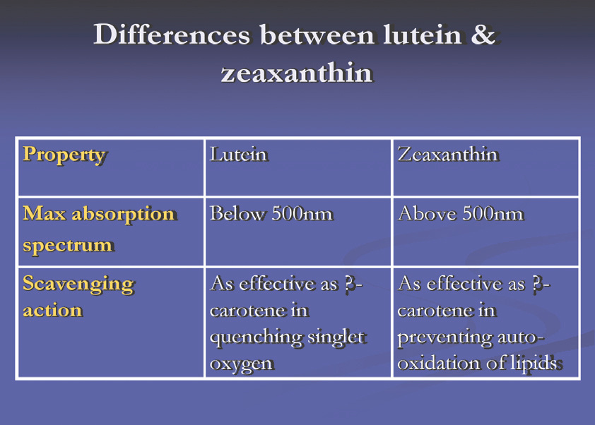
Figure 11, highlights the differences between L & Z
Functions of the xanthophylls:
The absorbance spectrum of MP peaks at 460nm (blue light), thereby protecting the macula from photo-oxidative damage. The fundamental means of blue light- induced damage is the photodynamic generation of free radicals from a toxic blend of oxygen and light (see part one). It is estimated that MP reduces the amount of blue light incident on the fovea by approximately 40%. This filtering function is especially important in young individuals (younger than 40 years), when the lens is virtually transparent to blue light.
MP also reduces chromatic aberration, with blue light primarily implicated in image degradation at the fovea.
Kirschfeld and later Khachik et al in 1997 demonstrated the antioxidant properties of carotenoids protecting the macula. Carotenois by virtue of their antioxidant activity may also play a role in regulating apoptosis (oxidative stress activates apopoptosis).
Furthermore carotenoids quench singlet oxygen by a physical mechanism, in which the excess energy of the singlet oxygen is transferred to the electron-rich structure of the carotenoid, without changing the structure of the carotenoid.
Singlet oxygen quenching depend on various parameters including; concentration and solubility in the tissue, oxygen partial pressure, ionic strength, viscosity, cell structural complexity and the presence of other redox-capable molecule or ions.
Carotenoids scavenge free radicals in two ways; firstly the free radical obtains its “missing” electron from the electron rich carotenoid, and secondly, the free radical adds itself to the carotenoid in an attempt to pair its single electron thus forming a covalent bond. Cell components (lipids, DNA and proteins) are thus spared from oxidative damage.
Vitamin E:
Vitamin E is recognized as one of the most important chain breaking antioxidants of cellular membranes. It consists of 8 tocopherols, the four most common being α-tocopherol (TP), β-tocopherol, y-tocopherol, and δ-tocopherol. Of these TP or a-tocopherol is not ids the most active and effective scavenger of free radicals, and the most predominant tocopherol in blood and tissues, including the retina. In addition this form appears to have the greatest nutritional significance; therefore vitamin E intake is often expressed in terms of milligrams of TP equivalent.
Vitamin E is absorbed from the intestines packaged in chylomicrons. It is delivered to the tissues via chylomicron, and then the remnants of chylomicrons are taken up by the liver that can export vitamin E in very-low-density lipoproteins. Due to its lipophyllic nature vitamin E accumulates in cellular membranes, fat deposits and lipoproteins. The major storage site of vitamin E is adipose tissue.
The vitamin E content of the RPE is 4-7x that of the neurosensory retina and rises with increasing age in response to oxidative stress. The concentrations in the tissues are very sensitive to the dietary intake of the vitamin.
Sources of TP, except fish, are largely limited to food sources rich in fat, such as vegetable oils, nuts, wholegrain, and green leafy vegetables. It is advisable to take TP supplements rather than increase high-fat foods in the diet.
Synthetic TP does not have the same biological potency as natural TP (only 67% as active as the natural form). The natural form is listed as d-alpha-tocopherol and the synthetic form as dl-alpha-tocopherol and is therefore easy to confuse.
Functions:
TP is known to be a physiological antioxidant that protects biological membranes from auto-oxidation. It is uniquely suited to intercept the peroxidation of PFA’S existing in cell membranes, preventing chain reactions in the membranes.
TP also protect lipids in LDL, as well as vitamin A from oxidation. Selenium, a micronutrient, complements the antioxidant function of vitamin E.
Vitamin C:
Ascorbic acid is a major water soluble antioxidant in the body with at least 300 metabolic functions. It is highly concentrated in all ocular tissues, especially the aqueous and lens (20x higher levels than the plasma). Vitamin C is entirely dietary dependent and cannot be manufactured by the body.
Sources include citric fruits such as strawberries, oranges, and grapefruit. As well as vegetables such as sweet red pepper, and tomatoes.
Best form of supplementation is esterified vitamin C (ester-C), which is non-acidic and enters the bloodstream 4x faster than any other form. Supplementation should be in divided doses combined with a bioflavanoid such as green tea. Remember that most dietary vitamin C is lost in the urine.
Functions:
The most clearly established role of vitamin C is collagen synthesis, where it acts as a cofactor in the hydroxylation of proline and lysine residues. Hence it is required for the maintenance of normal connective tissue, and wound healing. Vitamin C is also a highly effective antioxidant in the extracellular fluid and plasma due to its water solubility, stability, and the fact that it can be transported, reabsorbed and recycled. Vitamin E and C are interrelated in their antioxidant capabilities. Vitamin C may regenerate the active form of vitamin E following the scavenging of free radicals by TP.
Minerals and trace elements:
Minerals and trace elements are inorganic substances, which are essential for the maintenance of homeostasis and physiological functioning of the human body. Each mineral is required in specific amounts, ranging from micrograms to grams per day, with the optimal balance being crucial for the survival of every cell in the organism. It is estimated that around 20 different minerals are required in the human diet and of these only 7 have a well established biological role. Minerals can be categorized as macrominerals, required in quantities larger than 100mg. Macrominerals include Calcium, Magnesium and Phosphorus, and microminerals or trace elements where the requirement is less than 100mg. Trace elements include Zinc (Zn), Copper (Cu), Iron, Manganese (Mn), Chromium, Selenium (Se), and Iodine.
Mineral and trace element deficiency is more common than vitamin deficiency, with the elderly more at risk due to a reduced dietary intake. Furthermore, a reduction in stomach acid also reduces the release of trace elements from their ingested foodstuffs, and hence a decrease in their absorption and bioavailability.
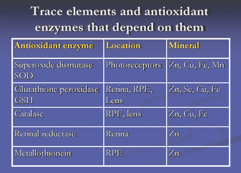
With ageing the antioxidant defense mechanisms effectiveness decline in part due to the decline of the bioavailability of some of the trace element cofactors. Increased oxidative stress has been proposed as the underlying mechanism for many age related diseases such as ARMD and cataract. (Part one). Figure 12 illustrates the relationship between trace elements and antioxidant enzymes.
The most important trace elements for purposes of this article are Zinc, Selenium, Manganese, and Copper.
Zinc (Zn):
Zinc is the most abundant trace element in the human body, with the total body content being approximately 2 grams. Tauber and Krauss (1943) first demonstrated the presence of Zn in the human eye. This was later confirmed by Galin et al who observed high concentrations of Zn in the retina-choroid complex (463mg/kg body weight). Recent studies have shown the maximum concentration in the RPE, 93.7% in macular area and 92.4% peripherally. Other tissues that contain Zn in descending order include the iris, choroid, sclera, vitreous, lens, cornea, and retina.
Sources include oysters, shellfish and red meat. Nuts and legumes are good plant sources. Of note is the fact that vegetarians need 50% more Zn than non vegetarians due to the high content of phylates in the diet which decrease Zn absorption from the digestive tract. Commercially Zn supplements are available as Zn acetate, gluconate, picolinate and sulphate. Zn picolinate is the most absorbable.
Tannins in red wine chelate metal cations in the digestive tract resulting in poor absorption of Zn. Large quantities of Zn in the diet can interfere with the bioavailability of Cu, therefore a 1:10 balance between Cu and Zn should be maintained with supplementation.
Function:
Zn is essential due to its role as cofactor in regulating the activity of specific Zn dependent enzyme systems. It is also involved in membrane stabilization as well as DNA and RNA synthesis. Zn also has a role in maintaining an intact immune system. In the eye its role in Vitamin A metabolism and as antioxidant deserves special mention.
Vitamin A metabolism
Zn plays a vital role in the transport and metabolism of vitamin A. In the retina Zn metallo-enzyme (retinol reductase), mediates the conversion of circulating retinol to active retinal. Retinal is then used for the synthesis of rhodopsin, which is responsible for scotopic vision.
Antioxidant properties
Zn stabilizes the cell membranes by protecting the cell against lipid peroxidation, which is particularly important in tissues such as the retina where high oxidative conditions prevail.
Zn stabilizes the structure of antioxidant enzymes such as SOD, which protect aerobic cells against the detrimental effects of superoxide radicals.
Zn also interfere with ROI binding and production, protecting the cell membrane against oxidative damage.
Taurine and Zn work synergistically to protect cell membranes from free radical damage by entrapping hydroxyl radicals. It also plays a role in apopotosis, which can be triggered by oxidative stress.
Selenium (Se):
The antioxidant enzyme glutathione peroxidase (GSH) is Se dependent. It acts in conjunction with vitamin E to protect cells against free radical damage by preventing lipid peroxidation. Se also regulates the effects of thyroid hormones on fat metabolism.
Sources include the Brazil nut, which is the most concentrated source containing 120µg of Se, seafood, meats, oats, brown rice, dairy products, fruit and vegetables, depending on the level of Se in the soil. Se is harmful if taken in excess.
Manganese (Mn) and Copper (Cu):
Mn functions primarily as a component of superoxide dismutases (SOD), which limits the damaging effects of the superoxide free radical from destroying cellular components. It is a vital catalyst in enzyme activity preventing calcification of soft tissue. Sources include dairy products, fish, meat, seafood, apples, avocado, and bananas.
Cu is present in SOD but its role in ocular tissue is not well understood. Cu works in conjunction with vitamin C and Zn to form elastin, an important skin protein. It is also essential for the formation of collagen.
Herbs:
Ginkgo Biloba:
This herb has powerful antioxidant properties that help increase blood circulation in the optic nerve, brain and retina. Ginkgo reduces the production of free radicals and plays a role in neurotransmitter metabolism. Studies have shown that retina and brain circulation is increased by up to 30%. Some studies have shown that ginkgo protects nerve cells during periods of ischemia and stimulates nerve regeneration. Ginkgo is used to treat dementia, vascular hearing loss in the elderly and erectile dysfunction secondary to atherosclerosis. It reduces vasospasm and serum viscosity and may increase memory retention. A study of 27 patients with bilateral normal tension glaucoma found ginkgo may improve preexisting visual field damage.
Ginkgo may interact with anticoagulants and anti-platelet agents causing bleeding due to the herb’s platelet-inhibiting properties. Exercise caution when using ginkgo with aspirin, Heparin, and Warfarin.
Bilberry extract:
The herb bilberry, a European relative of the American Blueberry, contains natural antioxidants that keep capillary walls strong and flexible. They also help to maintain the flexibility of the walls of red blood cells and alow them to pass through the capillaries better. It contains anthocyanidins or flavonoid pigments which occur in the cell vacuoles of plants and are responsible for the red, blue and purple colors. The flavonoid pigments help lower blood pressure, prevent clotting and enhance blood supply to the nervous system. It is estimated that anthocyanidins provide up to 50x the antioxidant protection of vitamin E and 10x the protection of vitamin C. The herb may help to support and strengthen collagen structures, inhibit the growth of bacteria in the urinary tract and have anti-inflammatory properties.
In the eye the antioxidant reservatol, and chemical compounds (anthocyanosides) prevent oxidative damage to the RPE cells and maintain rhodopsin levels required for night vision by the rods respectively.
| Nutrient | Function | RDA for Adults | Safe upper limit |
|---|---|---|---|
| Vitamin A | Maintenance of Skin, vision, lining of intestine, lungs, urinary tract. Helps protect against infection. | 1mg /day RE *men 0.8mg /day RE woman 700 -900µg, 1300µg for pregnant women | 3000g retinol, do not exceed 5000 IU in pregnant women |
| β-carotene | Major source of vitamin A in the body. Potent antioxidant properties | 4000 IU – 25000IU/day with lutein and zeazanthin | |
| Lutein & Zeazanthin MP | MP reduce incidence of blue light on macula, protect macula from photo-oxidative damage, quench singlet oxygen, scavenge free radicals | L 10mg/day and Z 2mg/day (AREDS 2) | |
| Vitamin E | Acts as an antioxidant. Protect vitamin A from oxidation | 10mg/day men, 8mg/day women, requirement increase with increased intake of PUFA’S | 800 IU or 540mg/day of d-alfa-tocopherol |
| Vitamin C | Formation and growth of bone, connective tissue, wound healing, function of blood vessels. Acts as antioxidant and helps body absorb iron. | 75mg/day women 90mg /daymen 35mg/day extra for smokers | 1000mg/day |
| Magnesium | Required for the formation of bone, teeth, normal nerve and muscle function, and the activation of enzymes | 300mg/day men 270mg/day women | 400mg/day |
| Selenium Se | Antioxidant with vitamin E. SOD** is Se dependent | 55µg/day (60 – 75µg) | 500µg/day |
| Zinc | Used to form many enzymes and insulin. Required for healthy skin, healing of wounds and growth | 15mg | 25mg |
| Manganese Mn | Component of SOD, prevents calcification of soft tissue | 2 -5mg/day | |
| Copper Cu | Component of SOD, forms elastin with vitamin C and Zinc. Essential in the formation of collagen | 0.5mg/day | |
| Gingko Biloba | Reduction in platelet aggregation and production of free radicals. Role in neurotransmitter metabolism. | Trials have used between 120 – 240mg/day | |
| Omega 3 fatty acids | Maintenance of cell membranes and production of prostaglandins which control inflammatory and immune responses | 6 -12g/day or 3 to 6% of total calories per day | |
| Omega 6 fatty acids | Maintenance of cell membranes, pro inflammatory | 6 -12g/day or 3 to 6% of total calories per day |
**SOD = Superoxidase dismutases
The next article in the series will deal with current research into nutrition related eye health, general rules to formulations, contraindications and side effects of supplements, drug interactions, and what to tell your patients about supplements.
References for this article can be obtained from the author via e-mail at d.booysen@pixie.co.za.
Part 3: Nutrition and Vitamin supplements, what do we tell our patients?
The first article in this series dealt with oxidative stress and its effect on the ocular tissue, and the second with the different nutrients, nutrient derivatives and herbs generally considered relevant to ocular health. If this has left you confused (as I am), hopefully this article will place things in perspective. I will briefly discuss the most important research into nutrition related eye health and the findings of these studies. Furthermore I will look at the general rules applicable to supplement formulations, the contraindications, adverse reactions, and the drug interactions which may occur with nutritional supplement use.
One of the problems I have experienced with supplement formulations is the confusion created by the different units used in the formulations. What is the relationship between IU or international units and mg? Surprisingly this was not easy to find and is more complex than expected.
IU measures the dose in terms of its biological effect rather than as a specific weight of pure substance. This was useful in the past for the following reasons.
- Sometimes it was not known exactly which chemical had the effect in the preparation
- Sometimes the amounts was so small it could not be assayed by the methods of the day
Since the 1950’s quantitative analysis of pharmacologicals became more precise and sensitive, allowing the substance responsible for an effect to be completely characterised by chemical and physical properties. The biological assay is no longer needed and the IU for that substance is discontinued. Some examples of old international units and their modern equivalents are:
- One IU of vitamin A = 0.3 micrograms (µg) of retinol or 0.60 micrograms (µg) of beta-carotene
- One retinol equivalent = 3.33 IU of retinol, or 10 IU of beta-carotene
- One IU of vitamin E = 0.91 milligrams (mg) of synthetic dl-alpha-tocopherol, or about 0.67 milligrams (mg) of d-alpha-topcopherol.
- One milligram (mg) = 1/1000 grams (g)
- One microgram (mcg or µg) = 1/1000000 grams (g)
What do the studies tell us?
Research into nutrition-related eye health is based mainly on outcomes from observational studies, and there is a need for randomized clinical trials proving the results seen in epidemiological data. In the mean time we have to look at the information we have available.
Age-related eye disease study (AREDS)
This study was initially conceived as a long term, multicentre, prospective study designed to asses the clinical course of, and risk factors for, ARMD and age-related cataract. In addition it was decided to incorporate a clinical trial of vitamin and mineral supplements for progression of ARMD and cataract, using high doses of these antioxidants.
In AREDS data were collected on potential risk factors as putatively determined by earlier laboratory and clinical studies. For ARMD, these risk factors included smoking, cardiovascular disease, hypertension, sunlight exposure and dietary intake of various micronutrients. For cataract, attention was directed towards educational status, smoking, diabetes, sunlight exposure, body mass index, drug use, oestrogen replacement therapy and dietary intake of various micronutrients.
Eleven retinal speciality clinics enrolled 4757 participants from 1992 to 1998, aged between 55 and 80 years. The average follow-up was 6.3 years with a range of 4 years for those participants that joined AREDS in 1997 (as the recruitment drew to a close) to 8 years for those who joined when the trial began in 1992.
Individuals fell into four categories.
- No ARMD, these participants had no drusen or only a few small drusen in one or both eyes
- Early ARMD, these participants had several small drusen or a few medium sized drusen, in one or both eyes
- Intermediate ARMD, these participants had many medium drusen or one or more large drusen, or areas of atrophy of the tissue outside the central macula in one or both eyes
- Advanced unilateral ARMD, these individuals had experienced the loss of some of the rods, cones and supporting tissue in the central retinal area (Dry ARMD), or had wet or neovascular changes in one eye only
All AREDS participants took 2 tablets bid for an average period of 6 -7 years. They did not have specific knowledge of what was in the medication, but they were informed that they would be randomly assigned to 1 of 4 different treatment groups:
- Zinc: 80mg zinc oxide plus 2mg cupric oxide
- Antioxidants alone; total daily dose of 500mg of vitamin C, 400 IU of vitamin E, and 15mg of beta-carotene
- Combination of antioxidants and zinc; 80mg of zinc oxide plus 2mg cupric oxide and a total daily dose of 500mg of vitamin C, 400 IU of vitamin E, and 15mg of beta-carotene
- Placebo with no active ingredients
Results
No difference in the development or progression of cataracts was seen for those individuals assigned to placebo as compared to those taking antioxidants.
The AREDS results showed no apparent benefit for the use of antioxidants plus zinc in individuals with early ARMD
The following table (1) summarises the results found in individuals with intermediate or advanced ARMD in one eye.
| Antioxidants + Zinc | Zinc alone | Antioxidants alone |
|---|---|---|
| – Reduced risk of developing advanced ARMD by approximately 25% | – Reduced risk of developing advanced ARMD by approximately 21% | – Reduced risk of developing advanced ARMD by approximately 17% |
| – Reduced risk of vision loss by approximately 19% | – Reduced risk of vision loss by approximately 11% | – Reduced risk of vision loss by approximately 10% |
Bressler et al estimated the potential public health benefit of the findings and reported that of the estimated 8 million individuals in the USA with monocular or binocular intermediate ARMD or monocular advanced ARMD, at least 300 000 (95% confidence interval, 158 000 – 487 000) people will avoid advanced ARMD on the AREDS formulation in a 5 year period.
The AREDS 2 study is a follow up of the original study, with the participants given a slightly altered formula containing, minerals, vitamins and 10mg Lutein, 2mg Zeazanthin , and/ or 1g omega 3 (DHA & EPA) per day. The purpose is to see if the supplements help slow the progression of ARMD to the advanced stages. Again the study is divided into 4 treatment groups as before and the plan is to enrol 4000 participants in categories 3 and 4.
- One group will be assigned to a placebo
- One to Lutein (L) 10mg + Zeazanthin (Z) 2mg
- One to omega 3
- One to a combination of L, Z and omega 3
In addition approximately half of the participants enrolled who choose to take the AREDS formulation will receive a new preparation without beta-carotene, and the dosage of zinc will be lowerd to 40mg. This will help to determine the benefit of beta-carotene in the original formulation. Results are pending.
Blue mountain study
This is an Australian study that used a questionnaire to evaluate the intake of 11 micronutrients, including Lutein, Zeazanthin, vitamin A, C via diet, zinc via diet, and supplements. The study found no significant associations or trends between dietary intake or dietary intake combined with zinc and the incidence of early ARMD, the 5 year incidence of ARMD, or the 5 year progression from category 1 or 2 to category 3 or 4. The study also reported on the association between vitamin and zinc supplementation and the prevalence of ARMD. They found no benefit for any type of supplement.
Lutein antioxidant supplementation trial (LAST)
The objective of the study was to determine whether nutritional supplementation with lutein (L), or L together with antioxidants, vitamins and minerals improved visual function and symptoms in atrophic ARMD. The study was a well organised prospective, 12 month, randomised, double masked, placebo controlled trial. 90 subjects with “dry AMD” were referred by ophthalmology and randomised into three groups.
- Group 1; Received 10mg non-esterified lutein per day
- oup 2; Received 10mg non-esterified lutein/ antioxidant/ vitamin/ mineral broad spectrum supplementation formula per day
- Group 3; Received a maltodextrin placebo
Outcomes were MPOD measured using heterochromic flicker, distance snellen equivalent visual acuity converted to logMAR, near visual acuity using low and high contrast SKILL test targets, contrast sensitivity, Amsler grid, and glare recovery symptoms.
At the end of the trial group 1 and 2’s MPOD increased by 0.09 log units, Snellen equivalent visual acuity improved by 5.4 letters for group 1 and 3.5 letters for group 2. Contrast sensitivity, Amsler grid, and glare recovery also improved for groups 1 and 2. Subjects in group 3 had no significant changes in any of the measured findings.
The study concluded that visual function improved with lutein or lutein together with other nutrients. This seems to prove that “dry AMD” is a nutrition responsive disorder and that improvements in visual function can be obtained over a short period of time. It is wise to consider this as a preliminary study due to the small number of subjects observed over a short time period.
The Aston randomised controlled trial (Aston RCT)
The purpose of the Aston RCT is to determine the effect of 18 months of daily lutein and antioxidant supplementation on measures of visual function in subjects with and without ARMD.
For inclusion, subjects have to present with no ocular pathology in one eye, or no ocular pathology other than “dry ARMD” in one eye. A grading system for cataract ( 1 = cortical, 2 = nuclear, 3 = posterior sub capsular )was also developed and subjects which had cataracts that precluded fundus photography were excluded.
Type 1 & 2 diabetics were excluded due to vitamin E’s affect on glucose tolerance, Subjects taking Warfarin were also excluded due to Zinc’s interference with its absorption and activity. Subjects with “wet ARMD” were also excluded.
The study formulation was:
- Lutein 6mg
- Vitamin A 750µg
- Vitamin C 250mg
- Vitamin E 34mg
- Zinc 10mg
- Copper 0.50mg
Outcome measures included, fundus photography, macular mapping test, glare recovery, visual acuity using Baily-Lovie logMAR charts, contrast sensitivity, and colour vision.
Results are pending?
Celtic age-related maculopathy arrestation study (CARMA)
The CARMA study is a multicentre study investigating the potential benefits of antioxidant supplements including lutei and zeazanthin on the course of ARMD. Recruitment started in July 2004 with preliminary results expected in 2007.
CARMA is a randomised controlled clinical trial of parallel-group design, where patients at high risk of developing advanced ARMD are invited to participate. Subjects are randomised to receive an antioxidant cocktail containing, vitamins C & E, minerals (Zn) and L + Z, or a placebo, and undergo tests of macular function at baseline and 6 monthly intervals. Differences between the groups in the rate of deterioration of visual function in response to antioxidant supplements, if observed, will strengthen the argument for screening for ARMD and the use of dietary modification with or without supplementation. This study will also provide insight into the potential role that antioxidants play in maintaining macular health. The plan is to enrol 500 patients who meet the inclusion criteria and the trial will last for 2 years with recruitment in the first 12 months. Clinical evaluations and procedures performed during the study include;
- Slit lamp bio microscopy of the anterior segment
- Dilated fundus examination
- Contrast sensitivity
- Colour photography
- Photopic interferometry
- Resonance Raman spectroscopy
- General physical examination
Results are pending?
General rules to prescribing formulations
Literally thousands of vitamin and supplement preparations are on the market and are used on a daily basis by our patients. So how do we decide on what to recommend for our patient eye health? Typically, when seeing a new patient, my case history not only includes finding out what medicines they are taking, but also learning what supplements they are currently using. You will be surprised how many patients are already taking some form of vitamin or mineral supplements, including speciality supplements to treat other problems.
As a rule I will discuss a general lifestyle and nutrition plan with my patients:
- Cease smoking immediately
- Use sunglasses and hats to reduce exposure to harmful light
- Eat at least 5 to 9 fresh organically grown fruits and vegetables per day
- This includes carrots, spinach, tomatoes, oranges and other green leaf vegetables
- Limit the intake of sweets and fats and use whole grain foods rather than refined foods
- Drink 6 to 8 glasses of water per day
Most patients who follow these guidelines probably require no additional supplementation. However, only about 9 to 10% of South Africans follow such guidelines and are therefore probably deficient in at least one third of essential vitamins and minerals. The evidence supporting the benefits of supplements are inconclusive as we have seen, but I believe that the vast majority of patients may benefit from some degree of supplementation. It is our duty to help them find a convenient program that addresses their needs.
One of the best researched vitamins is vitamin E. A good rule is to check the vitamin E content of a supplement, generally if it is in the right form (d-alpha-tocopherol) and concentration (200-400mg), the supplement typically has the other vitamins and minerals in the correct forms and amounts. Synthetic vitamins, although chemically “identical” to the natural form, are usually less efficient. In some instances, the synthetic form incorrectly binds to the cell receptor and blocks other reactions from occurring. This hinders cell function and can cause an increase in oxidative reactions, creating a “pro-oxidant” environment .
It is also a good idea to use a bid dose because of the water soluble vitamins (B complex and C) normally contained in the formulations which are excreted in the urine just 12 hours after ingestion. Stress to patients to look for supplements in capsule form and not tablet form as the process involved in tableting raises the internal temperature to several thousand degrees centigrade, causing the active ingredients to loose significant amounts of potency. Also look for supplements rich in amino acid chelates as they enhance bioavailability and reduce side effects when ionic salt forms of the minerals are used. Patients can take up to three times the RDA of B vitamins (RDA B2 = 1.1 -1.3mg, B6 = 1.5-1.7mg, B12 = 2.4µg). Vitamin C dose is 500 to 1000mg per day (RDA 75mg for women and 90mg for men with extra 35mg for smokers). Lutein and Zeazanthin can be taken in a 5 – 6mg divided dose per day. Ginkgo Biloba leaf extract is usually given in doses equalling 100 – 200mg per day. I also recommend Omega 3 fish oil that has an adequate amount of DHA (at least 120mg per 1000mg of fish oil) up to 1500mg per day. Be sure that the fish oil is emulsified to aid in absorption. Vitamin A dose is 500 -1000IU and beta carotene 4000IU. Dosage of Bilberry extract is 60 – 100mg per day, and Zinc 40mg per day with Copper 1.0mg per day.
The following is a formula I like using and have developed with a nutritionist. Recommended dosage, 1 -2 capsules bid with food. (Table 2)
| Ingredients | Per Capsule |
|---|---|
| Vitamin A | 10mg |
| Vitamin B1 | 10mg |
| Vitamin B2 | 5mg |
| Vitamin C | 300mg |
| Vitamin E | 54mg |
| Bilberry Extract | 40mg |
| Niacin | 20mg |
| Beta-Carotene | 15mg |
| Selenium | 0.2mg |
| Zeazanthin | 10mg |
| Ginkgo Biloba | 10mg |
| Lutein | 6mg |
| Zinc | 2mg |
| Copper Lyssinate | 2mg |
Contraindications and adverse reactions
“Generally, unless vitamins and supplements are taken in toxic doses the are harmless” This statement is not entirely true, but the risk of side effects from nutritional supplements is considerably lower than from over the counter or prescription drugs. The following will highlight the possible contraindications and adverse reactions for those nutrients reported to be beneficial to ocular health.
Provitamin A
Beta carotene was promoted as the preferred source of vitamin A due to the fact that it virtually has no adverse effects. However, doses of 20mg/day alone or in combination with alpha tocopherol (vitamin E) and 30mg/day in combination with retinyl palmitate (vitamin A) have been associated with an increased risk of lung cancer in smokers and those previously exposed to high levels of asbestos. Table 3.
| Study | Study subjects | Beta carotene | Outcome |
|---|---|---|---|
| ATBC (Alpha tocopherol/ beta carotene cancer prevention study) | Heavy Smokers, 50 to 70 years old | 20mg/day | 20% more lung cancer if beta carotene treated. No effect on cardiovascular disease |
| CARET trial | 20000 men and women. Smokers and/ or asbestos exposed | 30mg/day | Trend to more lung cancer and cardiovascular disease if beta carotene treated |
| Physicians health study | 20000 male physicians, 11% smokers, 40% past smokers, 90% non smokers | 50mg every other day | No increase or decrease in cancer or cardiovascular disease |
The evidence seems to indicate that supplemental beta carotene is not going to give any benefit to the individual in terms of cancer prevention or cardiovascular disease. In smokers, there may be harmful effects of beta carotene when it is broken down to vitamin A. This may be due to the fact that sometimes the beta carotene may break down into products that look like vitamin A and the cell is fooled, blocking the action of vitamin A or acting as a tumour-promoting agent. After all, smokers tend to be consumers of alcohol resulting in unhealthy liver function preventing detoxification of these vitamins, resulting in increased rates of cancer. It is also worth noting that the studies used synthetic vitamins in large doses and we know that a diet that provides these nutrients in the form of intact foods or natural form is more effective for disease prevention.
Beta carotene, more than 30mg per day can cause hypercaretonemia or reversible yellowing of the skin
Preformed vitamin A
Retinol can cause lower bone mineral density and increased risk of oesteoporotic fracture. Studies suggest that bone reabsorption is stimulated by vitamin A and also that vitamin A toxicity decreases bone formation. Vitamin A supplements have also been shown to increase the tendency to abnormal bleeding, which may indicate that it interacts with vitamin K required for production of coagulation proteins.
Hypervitaminosis A with symptoms of nausea, vomiting, increased cerebrospinal fluid pressure, headaches, blurred vision and poor muscle co-ordination can result in doses of vitamin A higher than 50000IU in adults and 10000IU in children. Symptoms usually disappear within 1 week of discontinuing the dose.
Vitamin A also reduces the activity of vitamin E by as much as 30%, but that vitamin A and D decrease the toxic effects of each other. Zinc may be specifically involved in mobilizing vitamin A from the liver to the circulation.
Vitamin C
Large doses of vitamin C are generally well tolerated but reduced bactericidal activity of leukocytes and insulin production may occur with routine doses of more than 1g over months or years. High doses may also cause stomach cramps, nausea, and diarrhoea.
Vitamin E
A randomised controlled trial showed that 400IU/day of vitamin E may have an adverse effect on the progress of retinitis pigmentosa. The hypothesis is that vitamin E may inhibit the absorption or transport of vitamin A. It may also exacerbate the effects of vitamin K deficiency and has been associated with an increase risk of haemorrhagic stroke and a reduced risk of ischemic stroke and ischemic heart disease. Anticoagulant (warfarin) use with vitamin E has been associated with abnormal bleeding and vitamin E appears to add to aspirin’s blood thinning effect.
Zinc
Zinc should be carefully used in type 1 diabetics as it increases glycosylation, or the addition of glucose to proteins, which is thought to be responsible for some of the clinical manifestations of type 1 diabetes.
Zinc interacts with copper by stimulating metallothionen levels of the intestinal wall. This binds to copper, preventing its absorption, which leads to copper deficiency and copper deficiency anaemia.
Zinc supplementation of 300mg/ day has been associated with impairment of immune function. Acute zinc toxicity has been reported with doses of 200mg or more and symptoms include headaches, abdominal cramps, loss of appetite, and vomiting.
Ginkgo Biloba
Ginkgo may thin the blood and should be avoided by patients on anticoagulant and antiplatelet drugs.
Drug interactions of ocular nutritional supplements
| Supplement | Drug | Explanation |
|---|---|---|
| Vitamin A | Anticonvulsants | Valproic acid may interfere with the body’s ability to handle vitamin A |
| Vitamin B6 | Folic acid | B6 may reduce the absorption or activity of folic acid |
| Vitamin C | Paracetamol | High doses of vitamin C may interfere with normal breakdown of this drug, and result in liver damaging accumulation of paracetamol |
| Vitamin C | Anticoagulants | Impaired blood coagulation time and interference with coagulation therapy |
| Vitamin E | Warfarin | 800IU daily vitamin E causes abnormal bleeding when added to the effects of warfarin |
| Vitamin E | Non-steroidal anti-inflammatory drugs | Vitamin E adds to aspirins blood thinning effects |
| Ginkgo Biloba | Anticonvulsants | Can prevent anticonvulsants from working as expected |
| Ginkgo Biloba | Warfarin | Reduces the ability of platelets to stick together, and add to blood thinning effects of warfarin |
| Ginkgo Biloba | Non-steroidal anti-inflammatory drugs | Increase the chance of abnormal bleeding when taken with aspirin |
| Magnesium | Diuretic | Amiloride may reduce urinary excretion of magnesium |
| Magnesium | Flouroquinolones Tetracycline’s Nitrofurantoin | Magnesium binds to these antibiotics greatly decreasing the absorption of the drug |
| Magnesium | Misoprostol | Magnesium can aggravate diarrhoea caused by misoprostol |
| Magnesium | Oral corticosteroids | Magnesium may interfere with the absorption of dexamethasone |
| Zinc | Fluoroquinolones | Zinc binds to these antibiotics greatly decreasing the absorption of the drugs |
Conclusion
The subject of nutritional supplements and their use in preventing or treating degenerative eye disease is controversial. However, some studies have clearly shown a benefit and others are currently in progress which should shed more light on this complex subject. I sincerely hope this series of articles have managed to shed some light on “what we know” and have encouraged my colleagues to explore the subject further.
Blaise Pascal, a French Philosopher, deduced that it was in his best interest to believe in God based on this argument. If you believe in God and there is a God, you will go to heaven. If you believe in God and there is no God, nothing will happen to you. If you don’t believe in God and there is no God, nothing will happen to you. But, if you don’t believe in God and there is a God, you will go to hell.
A similar argument can be made for nutritional supplements; therefore I recommend it to all my patients.
References:
- Nutritional and environmental influences on the eye, edited by Allen Taylor, CRC press
- Nutrition and the eye, a practical approach, edited by Frank Eperjesi and Stephen Beatty, Butterworth Heineman Elsevier
- Biochemistry of the eye, 2nd edition, David R. Whikehart, Butterworth Heineman
- Prescription for nutritional healing, third edition, Phyllis A. Balch, James A. Balch, Avery
- Biochemistry, International edition, Mary K. Campbell, Saunders college publishing
- The Merck Manual, 16th edition, MSD
- Concise Physiology, HS Meij, DH van Papendorp, Department of physiology UOFS
- The seeds of good health, Montgomery Vickers, Review of Optometry, Online
- Vitamins and age related macular degeneration, Shantan Reddy, Howard F. Fine, July/August 2006, Eyecare Africa
- What we know about AMD and Nutrition, Stephen G. Pratt, Review of Ophthalmology, Online
- The Nutrition Connection: The news on diet and eye diseases, Jefferey S. Eisenberg, Review of optometry, Online
- Can nutritional supplements benefit ocular health, Judith Springer Riddle, Review of Ophthalmology, Online
- What we know about AMD and nutrition, Steven G Pratt, Review of Ophthalmology, Online
- What we know about vitamins, Mark Abelson, Review of Ophthalmology, Online
- Can vitamin A make a comeback?, Mark Abelson, Review of Ophthalmology, Online
- MDs prescribe vitamins with reservation, Daniel Del Collo, Review of Ophthalmology, Online
- AMD and nutrition: The missing message, Christopher Kent, Review of Ophthalmology, Online
- New information about beta-carotene and smoking, Dan Roberts, mdsupport.org/library/betacarotene.html
- What to tell your patients about nutritional supplements, August L. Reader, Review of Ophthalmology, Online
- Are vitamins good medicine?, Paul Adjamian, Review of Optometry, Online
- More on self supplementation, Paul Adjamian, Review of Optometry, online
- International units, Pharmacology, www.sizes.com/units/international_units.htm
- One solution in the battle against dry eye: Nutrition, Jeffrey Anshel, Eyecare Africa, May/June 2006, p 26-30
- Retina, How lutein, laser therapy and steroid injections may help prevent vision loss from AMD, Mark T. Dunbar, Review of Optometry, Online
- AMD for the general ophthalmologist, Mary Elizabeth Hartnett, Review of Ophthalmology, Online
- Where do lutein and zeazanthin fit?, Paul Bernstein, Interview, Eyecare Africa, May/June 2006, p 26-27
- Supplements reduce risk of vision loss from AMD, Research review, Review of Ophthalmology, Online
- The effect of vitamin E and beta-carotene on the incidence of lung cancer and other cancers in smokers, Albanes et al, The new England Journal of Medicine, Vol 330, Number 15, April 14, 1994
- Vitamins and minerals: The results of the AREDS study, Kitty Sackett, Susan Bressler, www.agingeye.com
- Nutrition connection: News on diet and eye diseases, Jeffrey Eisenberg, Review of optometry, Online
- Clinical trial finds antioxidants and zinc beneficial in reducing risk of severe AMD, Tom Hoglund, www.mdssupport.org/library/study.html
- Nutritional supplements: Too much of a good thing, August L. Reader, Review of ophthalmology, Online
- Vitamin supplementation, what to tell your patients, Michael Beime, Review of Ophthalmology, Online
- Nutrition facts: Diet, Science and the eye, Stuart Richter, Review of Optometry, Online
- Protect your patients from vitamin toxicity, Steven G. Pratt, Review of Ophthalmology, Online
- Supplements reduce the risk of vision loss from AMD, Research review, Review of ophthalmology, Online
- AMD and the promise of AREDS, Mark Abelson, Review of ophthalmology, Online
- More thoughts on AREDS, AMD, Ann M. Hoscheit, Review of optometry, Online
- A closer look at AREDS and AMD patients, Carl Regillo, Emmet T. Cunningham, Review of ophthalmology, online
- Eye problems and eating disorders, Joseph P. Shovlin, Review of ophthalmology, Online
- Vitamin supplements – Too much of a good thing? Robert M. Russell, Joel Mason, www.thedoctorwillseeyounow.com/articles/nutrition/supplements_8
- Vitamins and age related macular degeneration, Shantan Reddy, July/August 2006, Eyecare Africa
- The mixed reactions of drug interactions, Bruce Onofrey, Review of Optometry, Online
- Can vitamins prevent cataracts?, Christopher Quinn, Review of optometry, online
- Zinc, antioxidants reduce AMD risk, Stephen F. Brint, Review of optometry, online
- Vitamin C cuts cataract risk in half, Review of optometry, Online