
Presented by Dr Dirk J Booysen at Contact Connect 2023
Content
- Introduction
- Inflammation
- Hypoxia
- Tear film changes
- Biofilms
- Keratitis
- Conclusion
Introduction
This is the story of contact lens related inflammation, hypoxia, tear film changes, biofilms, infection and their relationship to scleral lens adverse effects
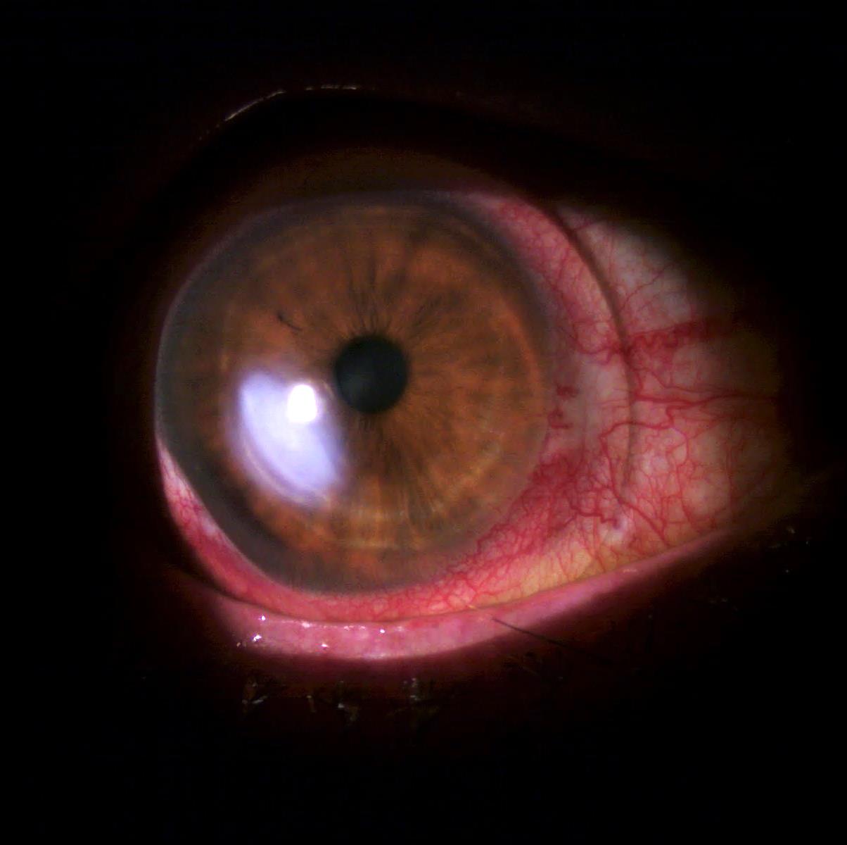
“All contact lens wear is intrinsically inflammatory”
Nathan Efron 2017
Add to this contact lens induced hypoxia, associated physiological challenge and bacteria, and the results may be catastrophic
Efron N. Contact lens wear is intrinsically inflammatory. Clin Exp Optom. 2017 Jan;100(1):3-19.
Inflammation
4 principal signs of inflammation (Celsus 30 BC – 45 AD)
5th sign later added by Galen (129 AD – 200 AD)
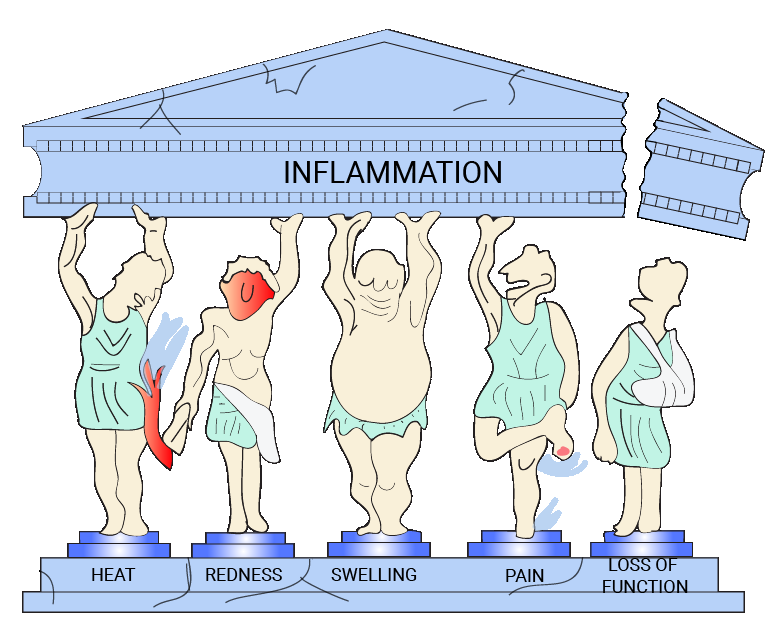
How does this relate to typical asymptomatic contact lens wear?
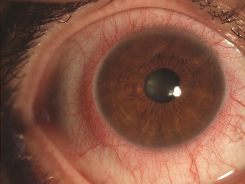
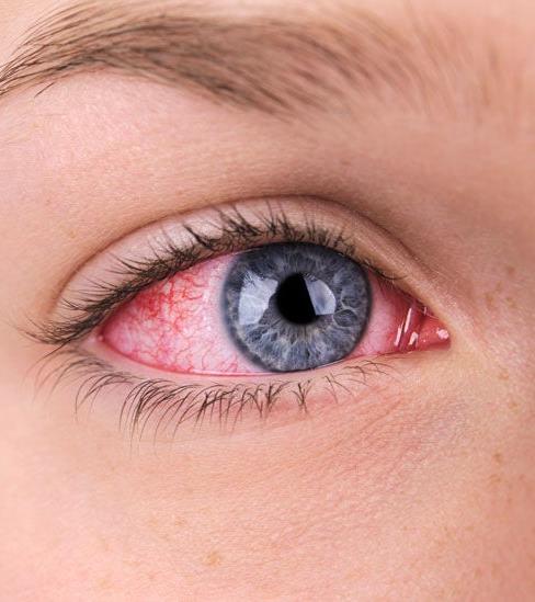
Rubor – hyperemia
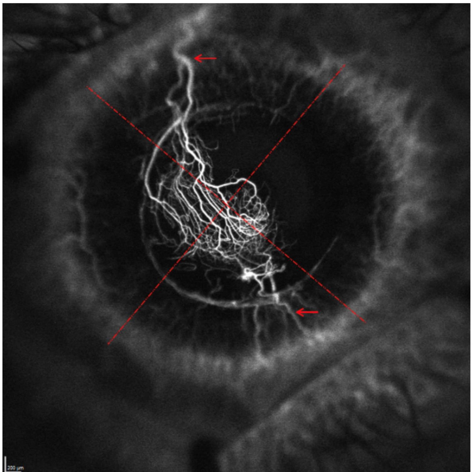
- The inflammatory response is mediated via tissue micro-vasculature
- The cornea is avascular – limbal hyperemia
- Limbal hyperemia is due to lens-induced hypoxia (Papas 2003) and hypoxia can be a direct stimulus to inflammation (Burki et al. 2014)
Calor – heat
- The normal ocular surface temperature is lower than the core body temperature (32.6 ± 1.24oC) – due to its exposed position as well as the cooling effect of evaporation (Yang and Zang, 2010)
- Corneal surface temperature increase with with contact lens wear especially toward corneal periphery (Martin and Fatt, 1986; Efron et al. 1989; Purslow et al. 2005)
- Pre-lens temperatures in soft lens wearers are cooler than the ocular surface without lenses, while the post-lens tear film temperatures are higher (Ooi et al. 2007, Craig et al. 2013)
- Lower water content lenses have less evaporation – surface temperatures are lower and post-lens tear film temperatures higher (Purslow et al. 2005)
- Conjunctival hyperemia and limbal hyperemia is significantly correlated with increased ocular surface temperature (Efron et al. 1988)
Tumor – corneal swelling
- Corneal swelling of 2-3% is common with scleral lens wear and in lenses that do not meet the oxygen transmissibility criteria of Holden and Mertz
- Hypoxia-induced osmotic corneal swelling can be considered an inflammatory response (Burki et al. 2014)
- What about IOP and corneal swelling with scleral lenses?
Dolor – pain
- Most contact lenses wearers will have some discomfort, especially at the end of the day (Craig et al., 2013, Efron, 2017)
- This is a leading cause of patients discontinuing lens wear (Craig et al., 2013)
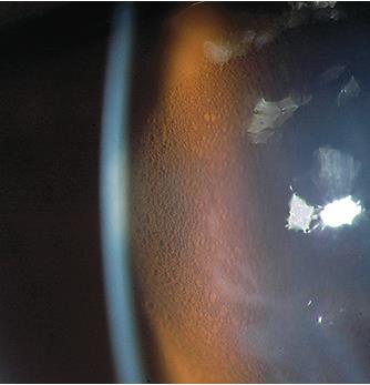
Functio laesa – loss of function
- More difficult to qualify in “asymptomatic lens wear” but if one considers the forced reduction in wearing time or discontinuation of lens wear due to discomfort or other issues an argument in loss of function of the affected tissue can be made (Efron, 2017)
What about “sub-clinical” or cellular, biochemical and molecular markers for inflammation?
- Tears contain many inflammatory mediators such as histamine, complement, arachidonic acid metabolites, substance P, cytokines (interferon and interleukins), tumor necrosis factor alpha, MMP 9 and epidermal factor (Dionne et al., 2016)
- “Although contact lens users were asymptomatic, changes in tear film levels of several important inflammatory mediators revealed that a chronic inflammatory process occurs during contact lens wear” (Yuksel Elgin et al. 2016)
- Stagnation of the tear film under the lens leads to inflammation due to retention of cells, microorganisms, and debris. This leads to an increase in the cytokine interleukin, MMP 9 and firbronectin, and a decrease in polymorphonuclear leucocyte recruitment and immunoglobulin A in the tear film (Stapleton et al., 2006)
Other inflammatory markers
- Among the cytokines and inflammatory mediators is prostaglandin – it’s role in the formation of neovascularization is well known (Efron, 2012)
- Plasma derived proteins, albumin and plasmin, are found in tear film of CL wearers – this indicates vascular leakage and albumin levels decrease when contact lenses are removed further indicating the presence of inflammatory cytokines in the tear film (Mann and Tige, 2013)
Inflammation Summary
- We can safely assume that during contact lens wear a chronic immune upregulation of the anterior ocular tissues occur (Efron, 2017 & Menkin, 1931))
- The eye is therefore in a steady state of readiness to suppress any challenge posed by chemical, hypoxic, traumatic, or infectious agents
Hypoxia
- As early as 1977, Fatt suggested that the contact lens and tear layer beneath or over them act as as series of resistors to oxygen transport
- The following formula can be used to obtain an estimate of the scleral lens system Dk/t
Dk/tscl = 1/(t1/Dk1) + (t2/Dk2)
t1 – lens thickness, Dk1 – lens Dk
t2 – post lens tear film thickness, Dk2 – tear film Dk usually 80 Dk units - Holden and Mertz (1983) established that 24 Dk/t units is needed to avoid hypoxia during daily wear followed by Harvitt and Bonanno (1999) criteria of 35 Dk/t units
- More recently Morgan (2010) indicated 20 Dk/t units centrally and 33 Dk/t units peripherally is necessary to alleviate hypoxia
Predicted values of Dk/t under the center of a scleral contact lens with Dk = 170 (Michaud, 2012)
| Dk=170 | Clearance (µm) | ||||||
|---|---|---|---|---|---|---|---|
| Lens thickness (µm) | 100 | 150 | 200 | 250 | 300 | 350 | 400 |
| 250 | 36.7 | 29.9 | 25.2 | 21,7 | 19.1 | 17.1 | 15.5 |
| 300 | 33.1 | 27.5 | 23.4 | 20.4 | 18.1 | 16.3 | 14.8 |
| 350 | 30.2 | 25.4 | 21.9 | 19.3 | 17.2 | 15.5 | 14.2 |
| 400 | 27.8 | 23.6 | 20.6 | 18.2 | 16.3 | 14.9 | 13.6 |
| 450 | 25.6 | 22.1 | 19.4 | 17.3 | 15.6 | 14.2 | 13.1 |
| 500 | 23.8 | 20.8 | 18.3 | 16.5 | 14.9 | 13.7 | 12.6 |
: satisfies HM criteria
: satisfies HM and HB criteria
In SA the most commonly use materials are Acuity 200 with Dk = 200, Optimum Extreme with Dk = 125 and Optimum Infinite with Dk = 180
Clearance in the periphery is less which affects the Dk/t positively
Apical Clearance and Lens Thickness – which is more important?
- If the scleral lens was thicker than 350 microns and the tear layer thickness exceeds 200 microns oxygen delivery to the cornea is adversely affected (Michaud, 2012)
- Large scleral lens wear results in 2-3% corneal oedema at the end of the wearing period if the lens and tear layer is thicker than 300 and 200 microns respectively (Van Der Worp, 2013)
- Amount of oxygen that reaches the cornea is inversely proportional to the amount of apical clearance (Michaud, 2012)
- Reducing lens thickness in modern high DK materials have little clinical benefit in reducing corneal oedema during open eye lens wear – only statistical difference obtained for 150 and 1200μm thickness comparison (Fisher D, Collins M, Vincent S, 2022)
- 0.45% of SCL wearers develop corneal oedema and 0.01% bullae (Schornack et al., 2016)
Minimizing hypoxia
- To minimize hypoxia induced corneal swelling with scleral lenses use: (Michaud et al., 2012)
- lenses with high Dk values >150
- maximal centre thickness of <250 microns or less
- and tear lens thickness not exceeding 200 microns
- Scleral lens materials should have a Dk of at least 125 with a thickness of 200 microns and a tear film thickness of 150 microns or less to meet oxygen tension of 55mmHg – which is considered the minimum critical barrier to avoid clinically significant hypoxia. (Compan et al., 2014)
- Corneal oxygen consumption would be higher after a patient has been wearing thick rigid gas-permeable lenses with higher corneal clearances (Jaynes et al., 2015)
- It is not inconceivable to expect a low-grade chronic hypoxic state with scleral lens wear that may promote oedema and neovascularization (Safvati et al., 2009)
- On average the cornea will swell by around 1.6-3% (Compan et al., 2014, Pullum and. Stapleton, 1997)
What about Tear Exchange Under a Scleral lens?
- The amount of tear exchange under a scleral lens is minimal and depends on the lens diameter, the larger the lens the longer it takes to replenish the fluid under the lens
- On average tear exchange for a 16mm scleral lens is limited to a rate of 0.2% per minute once the lens settles on the eye (Vance et al., 2015)
- Based on this rate, it would take >8 hours to replenish the fluid under the average scleral lens
- Accumulation of metabolic by products can contribute to the onset of adverse events primarily by altering the epithelial barrier function (Muntz et al., 2015)
- Without good tear exchange, the incidence of microbial keratitis (MK), contact lens acute red eye (CLARE), and contact lens-induced corneal oedema increases (Sonsino, 2015)
Tear film changes
Blink
- Higher than normal percentage of incomplete blinks (Van der Worp, 2008)
- Increased inter-blink interval (Craig et al., 2013)
Tears
- Post-lens tear stagnation leads – increased inflammation due to retention of cells, microorganisms and debris (Stapleton et al., 2006)
- Aqueous layer is split in two – 2-6µm thick pre-lens tear film – disrupted lipid layer (Craig et al., 2013, Nong et al., 2010, Stapleton et al., 2006)
- The thin pre-lens tear film influenced by lens diameter, lens type, fit, surface chemistry and wearing schedule (Stapleton et al., 2006)
- Lipid layer directly interact with the lens surface causing poor wetting and deposits (Yokoi et al., 2008)
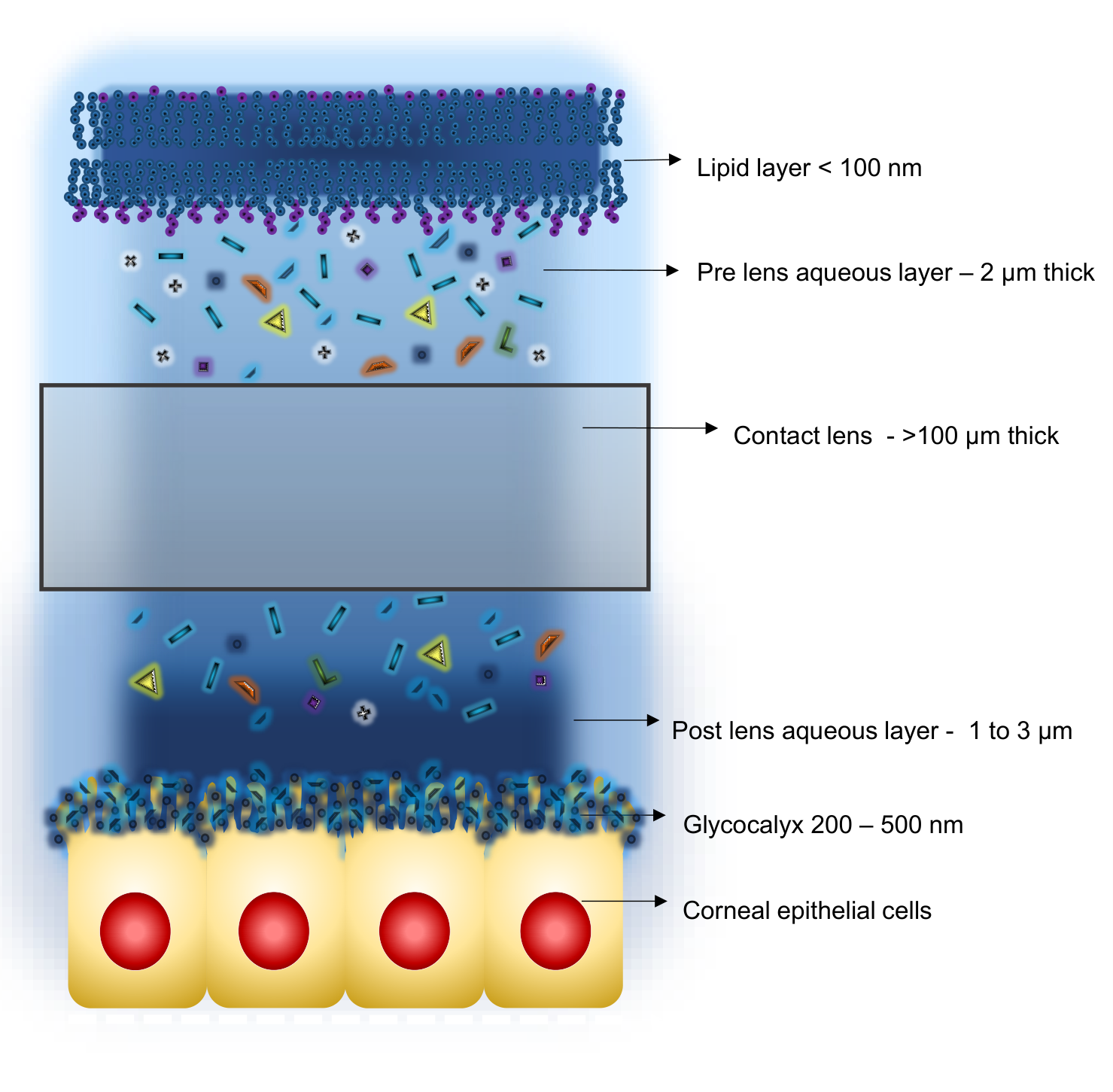
- Clinically pre-lens lipid layer is absent in RGP lenses – reducing TBUT to around 2 – 3 seconds and 5-6 seconds with soft lenses (Craig et al., 2013)
- Disruption of the lipid layer cause increased tear film evaporation – 1.2 – 1.6x with contact lenses in situ (Guilln and Maissa, 2008, Refojo, 1991)
- Increased evaporation increases tear osmolarity, discomfort and dryness (Craig et al., 2013, Guillion and Maissa., 2008, Refojo, 1991)
- Post-lens tear film temperature is higher in contact lens wear (Craig et al., 2013, Ooi., et al., 2007, Purslow et al., 2005)
- Tear turnover rate decreases in CL wear from an average of 15.5%/minute to 12.4%/minute in hydrogel and 13.2%/minute in silicone hydrogel wear (Occhipinti et al., 1988, Kok et al., 1992, Craig et. Al., 2013)
- Tear meniscus volume decreases from 1.5µl to 1µl contributing to discomfort (Palakuru et al., 2007, Chen. Et al., 2010)
- Tear pH decrease in CL wear and especially the post lens tear film becomes more acidic due to CO2 accumulation (Chen and Maurice, 1990, Craig et al., 2013)
- Tear protein attaches on the lens surface and in the matrix, denatures and is then not recognized as “self” – linked to biofilms and CLPC (Mann and Tige, 2013)
- CL wear and cleaning solutions affect the amount of secreted mucins and damage the glycocalyx formed by the transmembrane mucins (Craig et al., 2013)
Biofilms
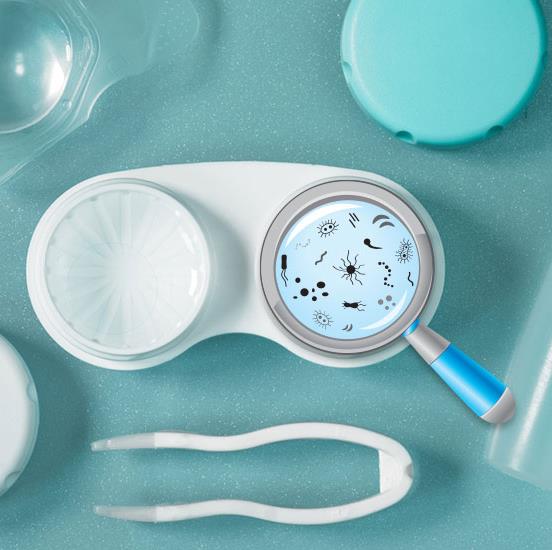
- 40-80% of bacteria on Earth can form biofilms (Flemming and Wuertz, 2019)
- Biofilms are complex surface attached communities of microorganisms held. Together by self-produced polymer matrixes composed of polysaccharides, secreted proteins, and extracellular DNA (Tremblay at al., 2013)
- Can be a single organism or multiple species including fungi, yeast, protozoa, bacteria and algae (Tomaras et al., 2003)
- Biofilm formation on the ocular surface is prevented by the sweeping action of the lids, flushing of the tears, lactoferrin, lysozyme, and probably most importantly mucin secretion from the goblet cells – protecting the epithelium (Kurpakus Wheater, Kernacki & Hazlett, 1999)
More about Biofilms
- Both S.epidermidis and S.aureus are excellent biofilm formers, especially S.epidermidis (Shokouhfard et al., 2015)
- P. aeruginosa were the most potent biofilm forming microorganisms on all kinds of CLs in every condition (Dosler et al., 2020)
- Most common bacteria causing biofilms on CL – P. aeruginosa, S. aureus, S. epidermidis, S. sacrophyctius, Klebsiella spp. (El-Ganiny et al., 2017)
- Anti-biofilm activities of MPSs were based on various factors, such as chemical ingredients and contact time of MPSs, the type of infectious agent, and especially the CL type and usage time (Dosler et al., 2020)
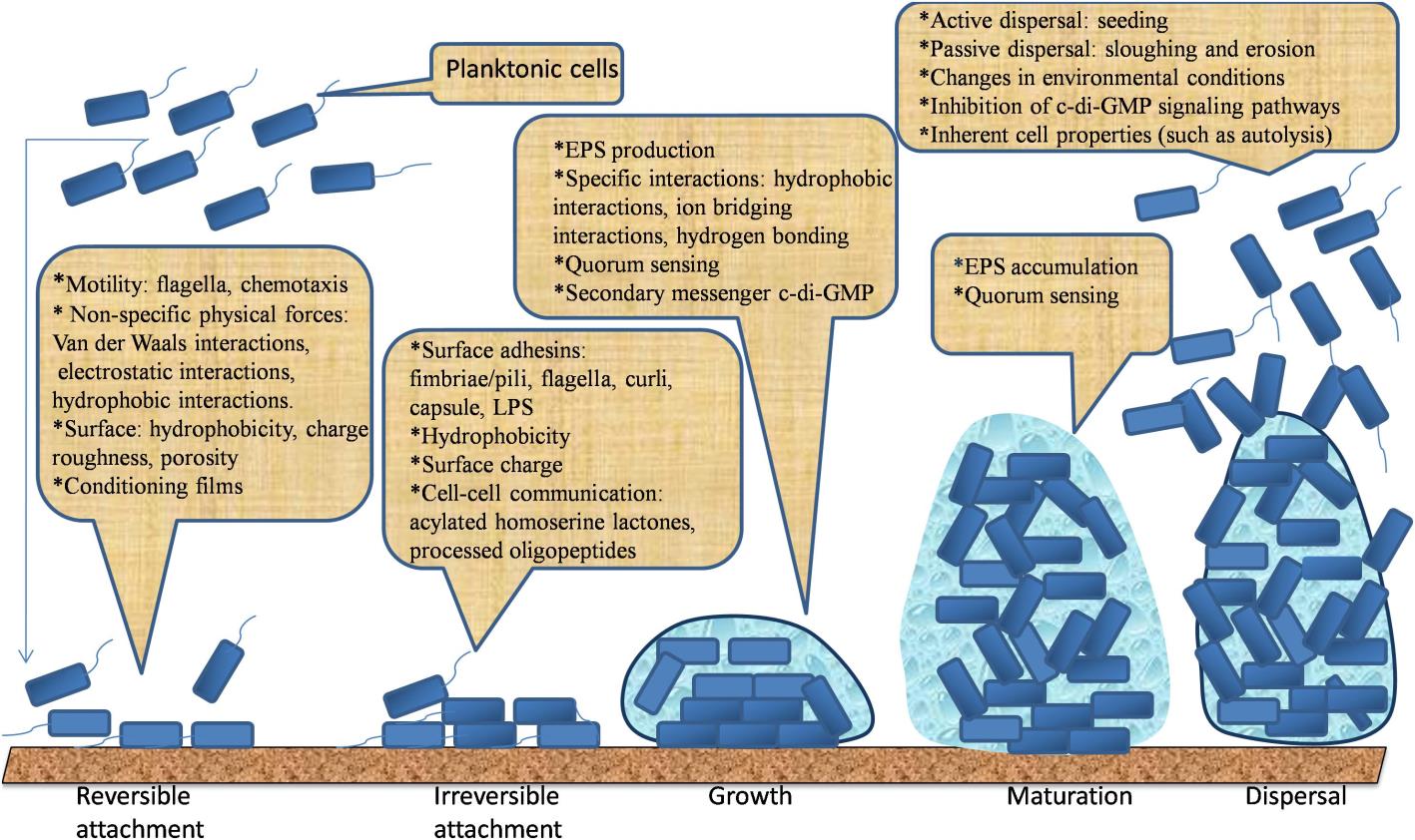
LPS – lipopolysaccharide
What makes the surface more prone to biofilm formation?
- Bacteria are primarily negatively charged and complex interactions involving electrostatic forces, Van Der Waals forces, hydrophobic interactions, steric forces play a role in biofilm formation and adhesion.
- In short, rough, hydrophobic materials with a positive charge are more susceptible to biofilm formation
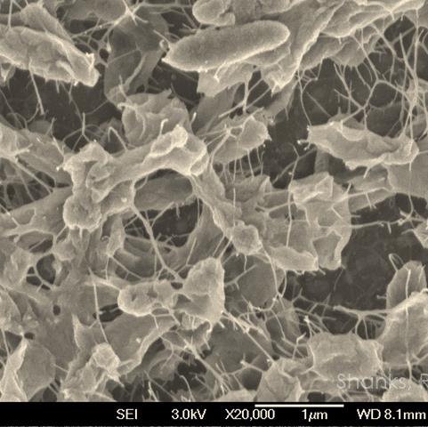
Biofilms on Contact Lenses
- Biofilm thickening starts much earlier in contact lens wearers
- The contact lens is itself an inert foreign body allowing bacteria to avoid host defence and get a head start, producing a very early adherent biofilm (Dosler et el. 2020)
- As previously mentioned, CL’ reduce blinking rate, alters the tear film as well as tear flow and exchange
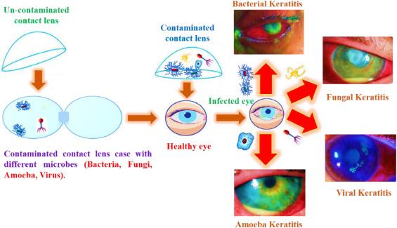
Corneal defense mechanisms
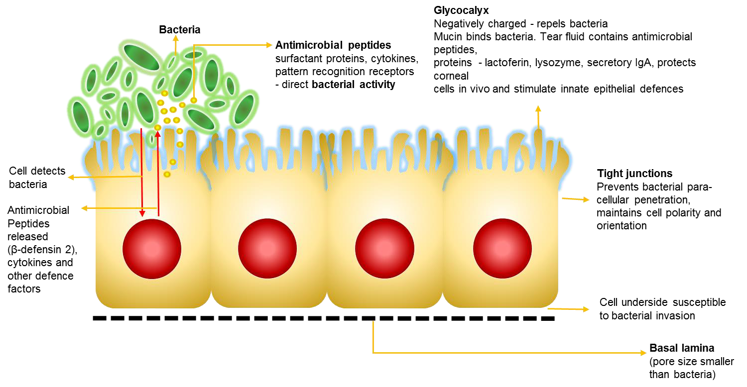
Three Critical Defense Layers prevent Infection
- Tears, Epithelium, Basal lamina
- Redundancy suggests all three may need to be compromised to get corneal infection
- “Infection follows changes to ocular surface biochemistry under a lens” (Fleizig)
Now that we have an understanding of the factors involved let’s have a look at some of the scleral lens adverse effects and the mechanism by which they occur
Common Scleral Lens Adverse Effects
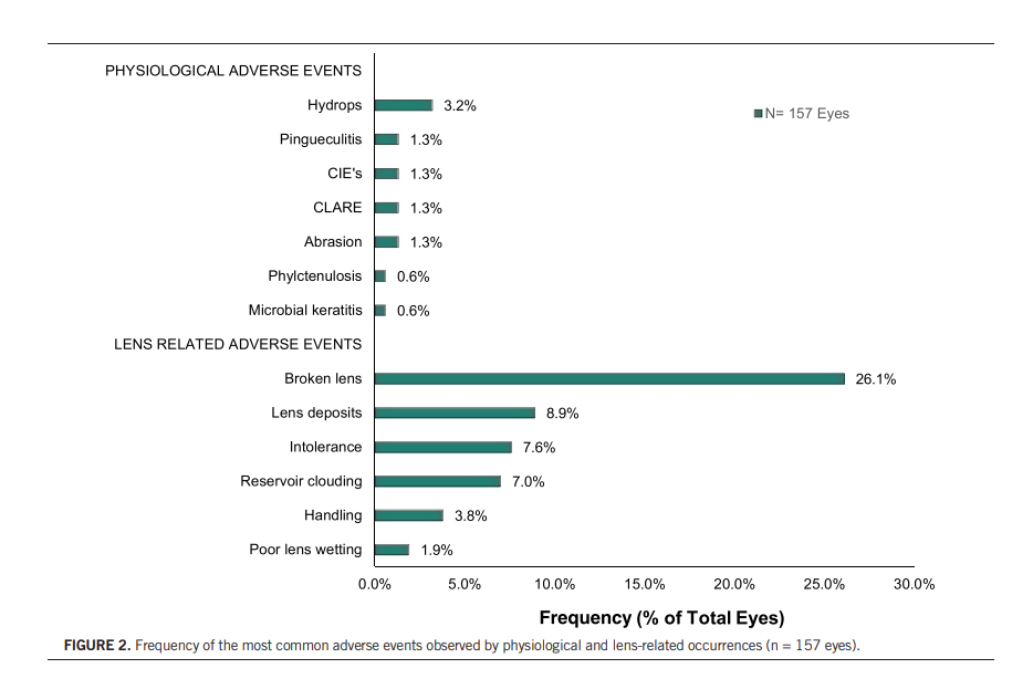
Daniel G. Fuller, Yueren Wang. Optom Vis Sci 2020; Vol 97(9)
10 most common SCL adverse effects
10 – Bubbles
9 – Conjunctival blanching
8 – Conjunctival prolapse
7 – Pain or discomfort
6 – Settling back
5 – Corneal staining
4 – Non-wetting and surface deposits
3 – Handling problems
2 – Conjunctival redness
1 – Misting under the lens or midday fogging
(Taylor S, Optician Online)
Conjunctival prolapse
- Caused by pressure or fluid forces generated behind a sealed scleral lens and occurs in locations of excessive limbal clearance(Fisher et al., 2021)
- More common in patients conjunctivochalasis and PMD
- Management
- Reduce excessive limbal clearance
- Reduce excessive central clearance
- Toric or asymmetrical peripheral design
- Considered benign and does not seem to affect cornea in the short term –long term can cause issues with neovascularization and conjunctivalisation (Fisher et al., 2021)
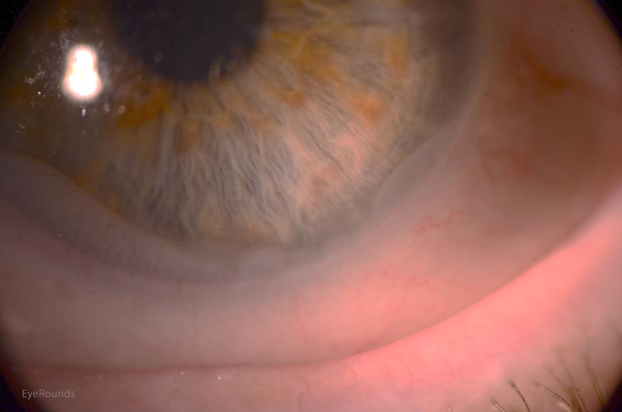
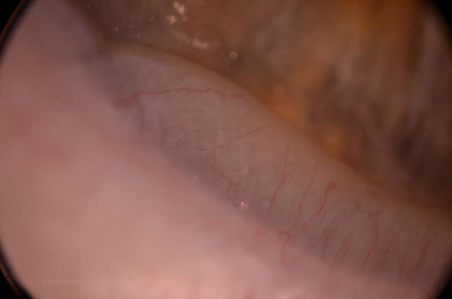
Surface soiling poorly wetting lenses
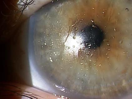
- One of the most challenging adverse effects to deal with with no obvious cause!
- Tear film is altered with lens wear, especially the lipid and mucin layers
- Conjunctival goblet cells affected by landing zone of the lens – mucin layer altered
- MGD, dry eye, blepharitis, atopy, hormonal, medication this list is long
- High DK/t, silicone-based materials – lipid deposits
- Hand lotions, make up on lens surface
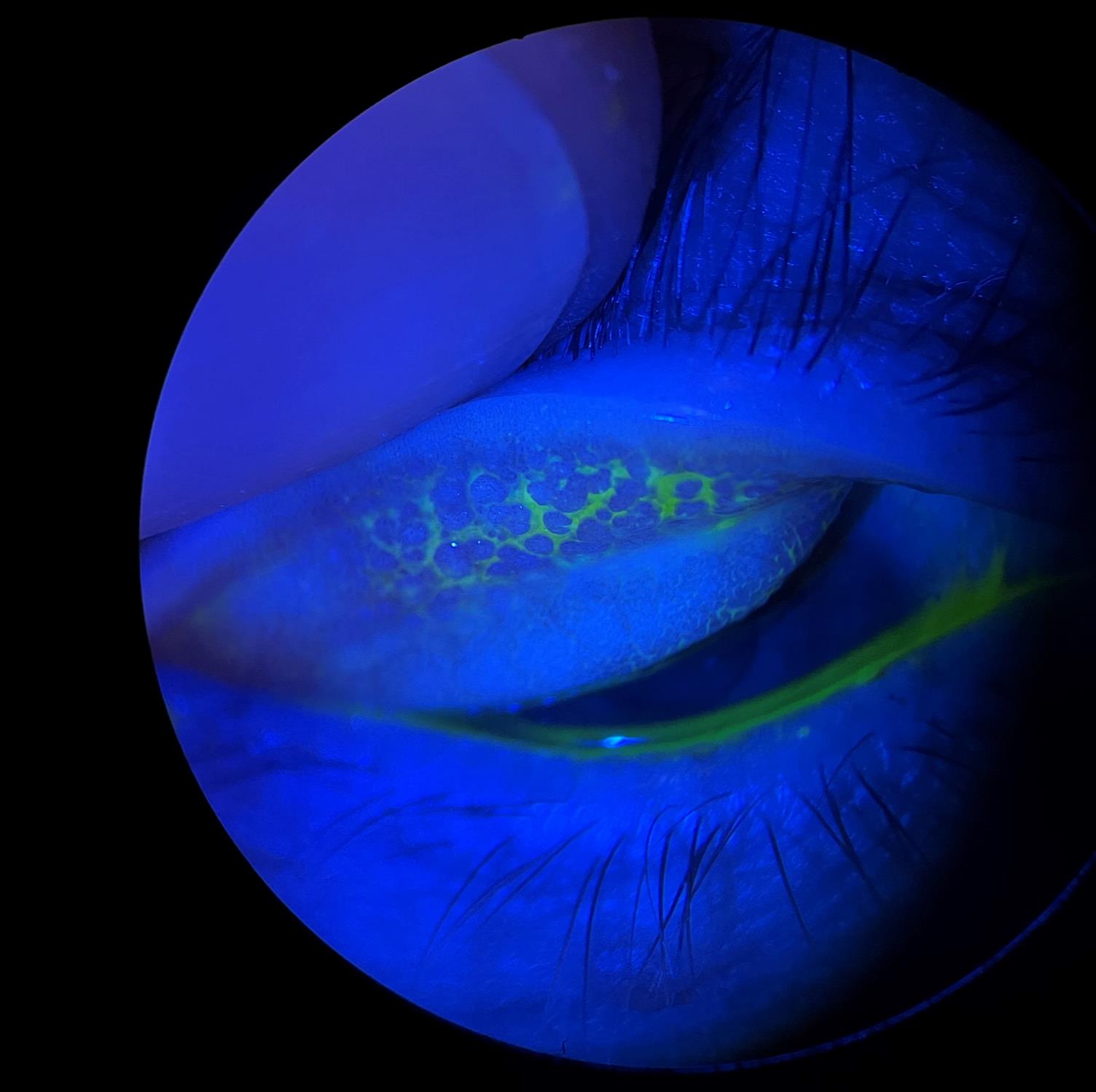
What about friction and lid wiper epitheliopathy?
- On average, most people blink around 15 to 20 times each minute, 900 – 1.200 times an hour or 14.400 – 19.200 times a day
- Abrasion, inflammation and poor spread of the tear film over the lens surface
- Frequent lubrication, debride the lid wiper, Hydra-PEG coatings
What about GPC or CLPC?
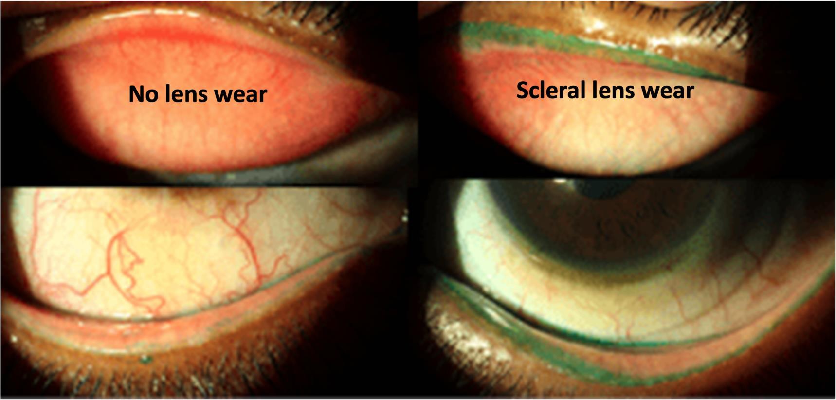
- SCL wear can be associated with GPC
- Particularly in lenses with overly flat superior haptics and excessive superior edge lift
- This causes lens discomfort (GPC), and increase mucous debris in the post-lens fluid reservoir (Johns et al, 2017)
Epithelial bogging
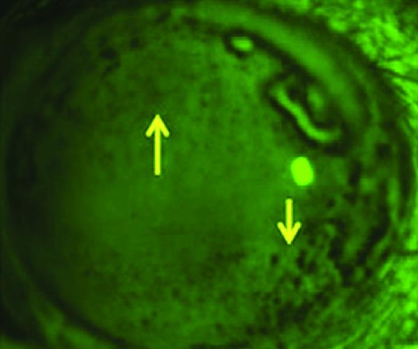
- Epithelium appears rough, irregular or waterlogged
- Exact aetiology unknown – possibly due to cells remaining in contact with the stagnant tear film between the lens and the eye (Walker et al., 2016)
- It may represent epithelial oedema, loss of glycocalyx layer leading to surface wetting problems, or an osmotic balance in the tear film (Walker et al., 2016)
- Interference with normal lid sweeping action over the epithelium may allow non-vital cells to build up (Walker et al., 2016)
Midday fogging
- Incidence 20-30% of all scleral lens wearers (Walker et al., 2016)
- Particulate matter in the post-lens tear film associated with blurred vision (Garracedo et al., 2017)
- Aetiology seems to be related to lens induced mechanical trauma to conjunctiva/limbus/cornea, hypoxic inflammation and meibomian gland lipids accumulating in the tear reservoir (Garracedo et al., 2017, McKinney et al., 2013)
- Corneal oedema due to hypoxia can also contribute
- Increased mucin production from rubbing the conjunctival tissue can also contribute
- Accumulation of protein and lipid on the lens surface (Barnett & Johns., 2018)
- Treatment consist of ruling out causes and adjusting the lens fit accordingly
- Midday fogging can be prevented or alleviated by using non-preserved saline mixed with thick preservative free artificial tears when inserting the lens (Walker et al., 2016)
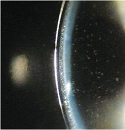
Bulbar and Limbal Hyperaemia
- Contact lens induced inflammation
- Mechanical trauma, irritation and compression
- Lens seal-off – no tear exchange and exposes cornea to toxic waste – common. With PRK, LASIK, RK or oblate corneas
- Infection
- Remedy:
- Check if lens is not excessively bearing on the circum limbal area
- Increase limbal clearance
- Loosen peripheral curves/ toric design
- Reduce diameter of the scleral lens
- Flatten and widen landing zone
- Preservative free solutions
- Treat infection and inflammation
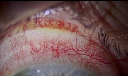
Neovascularization
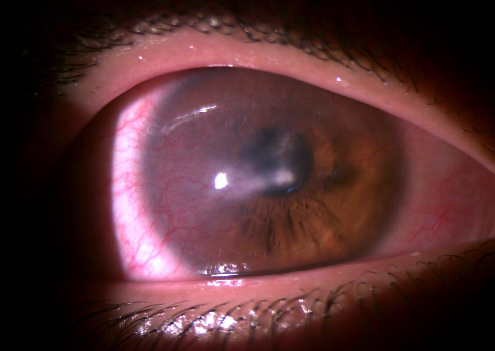
- Hypoxia leads to corneal oedema and stromal softening.
- Mechanical trauma and hypoxia leads to inflammation and the release of cytokines and prostaglandins which are angiogenic.
- Inflammatory cells migrate into the cornea further releasing vasostimulating agents initiating new vessel growth.
- The cellular distress signal directly triggers vascular endothelial mesenchyme forming: – endothelial cells, pericytes, fibroblasts and the smooth muscle of new blood vessels
- 0.28% of SCL wearers (Schornack et al., 2016)
Keratitis
Why do contact lenses cause infection?
Hypoxia is not necessarily required but could play a role via metabolism thereby influencing the corneal defence mechanisms
- Defensin production
- Epithelial cell polarity, tight junctions
- Cell sloughing and cell replacement
- Tear production
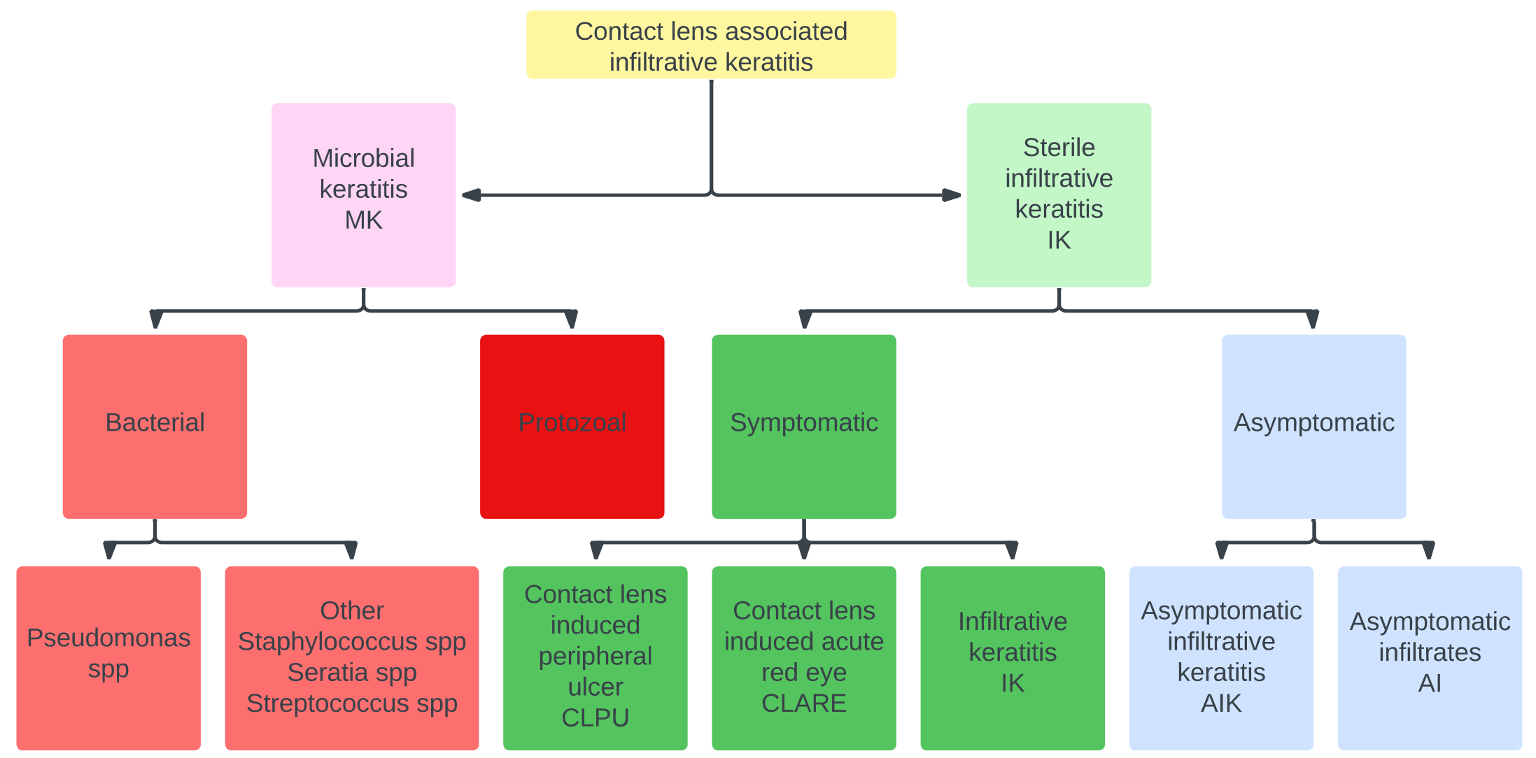
IK is rare with scleral lens wear ~ 0.2% (Walker et al., 2016 and Schornack et al., 2016)
MK even lower risk ~ 0.1% (Schornack et al., 2016)
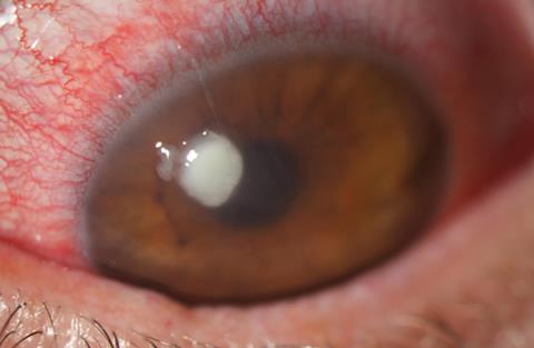
Classification of CL associated Infiltrative Keratitis
Conclusion
SCL clinicians feel like Super-Hero’s, however, even the likes of Superman has a bad day every now and then!
- Your specialized training should equip you with the knowledge to deal with all SCL associated adverse effects
- Deal with them timeously and appropriately within your comfort zone and scope
- Always remember
“When in doubt send it out”