

Dip Optom FOA(SA), D Optom (Aston), FC Optom(UK), TMOD (PCO), CAS Neuro(NECO), FSLS, FISCLS, PGCOT (Suny)
SAOA July 2024
dirk@dirkbooysen.co.za
Content
- Review of vitreous anatomy/biochemistry
- Review of the vitreous physiology and function
- Vitreoretinal attachments
- Examination of the vitreous
- Symptoms and signs of trouble
- Vitreous degeneration/detachment/retinal detachment
- Posterior hyaloid membrane detachment, vitreomacular adhesion/traction, epiretinal membranes, cystoid macular oedema and macular holes
- Vitreous opacities
- Conclusion

Introduction
- The vitreous body, ominously called “vile jelly” by Shakespeare, has been of therapeutic interest since ancient times
- According to Eber’s papyrus (1400 BC), the Egyptians thought that the inner part of the eye communicated by a canalicular system with the inner ear cavities and the lacrimal cavity
- The Egyptians hoped to restore lost vision by replacing the inner part of the eye with a lyophilized mixture injected into the inner ear
Ancient Egyptian
- Eber’s papyrus states that the best mixture to replace the vitreous was also a remedy for total blindness:
- “extract of pig’s eye humour 1 part,
- genuine Galenos 1 part,
- yellow ochre 1 part,
- fermented honey 1 part”
- Today, the vitreous is known to provide an optically clear medium, a mechanical buffer, and a nutrient tissue

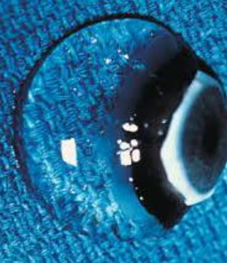
Vitreous anatomy
- Ophthalmoscopically transparent but structure can be observed with slit lamp
- Rule of “4’s”
- 4 grams
- 4 cc
- Occupies 4/5 of the globe
- 4 x as viscous as water at birth
- Consistency similar too the albumin of an egg
Substructure of the vitreous
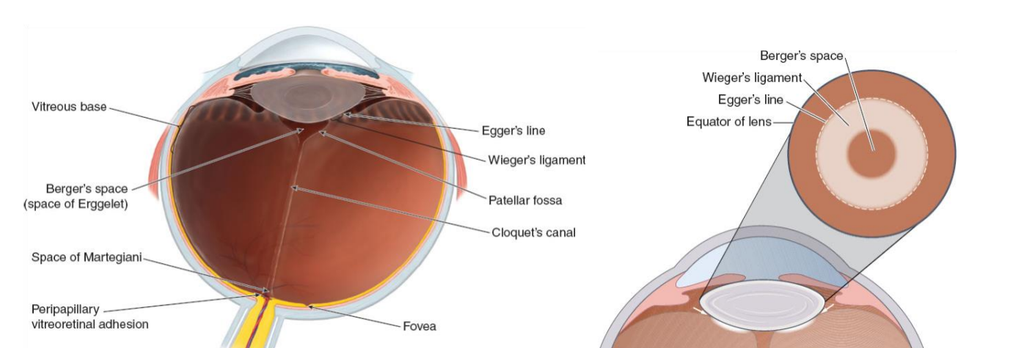
Anatomical openings, spaces an attachments play an important role in the development of pathology
Vitreous biochemistry
- 99% water, rest solids
- Acts as a viscous gel or interconnected meshwork
- Gel structure contains long, thick, nonbranching, mostly type 2 collagen fibrils suspended in a network of hyaluronic acid (HA) – type IV collagen – crosslinks
- Proteins (fibrillin & opsin), amino acids, vitamin C (ascorbic acid), and cells (hyalocytes and fibroblasts) are also found
- Worst suggest the adult vitreous body is composed of a number of cisterns
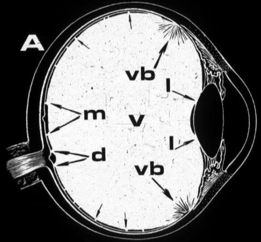
Vitreoretinal interface
- The cortex is composed of two parts;
- anterior cortex/anterior hyaloid face, begins about 1.5 mm anterior to the ora serrata
- Daicker likened this structure to Velcro – this explains the strong vitreoretinal attachment at this site
- posterior cortex/hyaloid – 100-110 µm thick and consists of densely packed collagen fibrils.
- Hogan demonstrated that just posterior to the ora, heavy bundles of vitreous fibrils attach to the basal lamina of retinal glial cells (Müller Cells)
- There is no cortex over the disk, and the cortex is thin over the macula and fibers are more spread out
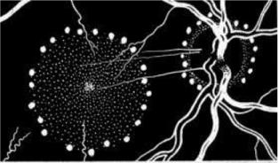
Vitreoretinal interface – Hyaloid and ILM
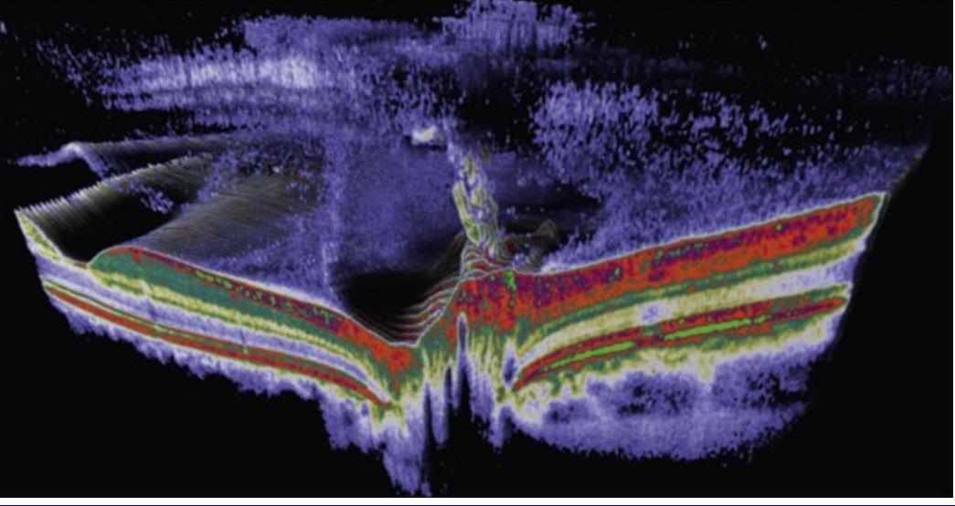
- The cortex/hyaloid is firmly attached to the ILM of the retina at the vitreous
base, around the disc (Weiss ring), at the vessels, and the foveola at a
diameter of 500 µm - The ILM is 1 – 3 µm thick, consisting
mainly of type IV collagen and proteoglycans. - The ILM can be considered the basal lamina of the Müller cells
- The only region not adjacent to a basal lamina/ILM is the anterior vitreous cortex annulus that is directly exposed to the zonule and the aqueous humour of the posterior chamber
- Balazs has stated that the basal lamina/ILM prevents the passage of molecules larger than 15-20 nm and proposed that the complex of the posterior vitreous cortex and ILM could thus act as a “molecular sieve”
Vitreoretinal interface – Hyaloid and ILM
- Reeser and Aaberg consider the posterior vitreous cortex as the “metabolic center” of the vitreous because of the presence of hyalocytes embedded within the cortex
- Hyalocytes synthesize HA, matrix glycosaminoglycans and glycoproteins
- Hyalocytes become phagocytic cells in response to noxious stimuli and inflammation
Clinical relevance
- The vitreous is most firmly attached at the vitreous base, the disk, and the macula, and over retinal blood vessels particularly in the fundus periphery
- With typical vitreous detachment, the vitreous detaches everywhere except where the stronger anatomical adhesions are present
- This may result in retinal detachment
- Once a PVD occurs, the normal holes that exist in the cortical vitreous – at the macula and disc, permit the liquefied vitreous to escape into the sub-hyaloid space.
- This collapses the vitreous cortex, accelerating its further detachment from the retina up to the posterior border of the vitreous base
Functions of the “silent gel”
Developmental function
- Growth of the eye must occur to exact specifications since the various tissues must bear precise geometric relations to one another if optic and photoreceptive functions are to be achieved
Optical functions
- Clarity
- The index of refraction of vitreous is 1.3349, nearly the same as that of aqueous
- Vitreous transmits 90% of light between 300 and 1400 nm and none above or below this range
- Accommodation
- Cramer proposed that the vitreous pushes against the lens to alter its shape
- These theories, however, largely ignored the influence of zonule relaxation and lens capsule elasticity
- In this context, it is interesting to consider that significant liquefaction of the vitreous begins at about the same age as the onset of presbyopia
Other functions
- Mechanical functions
- The viscoelastic characteristics are well-suited for a shockabsorption function
- Considering the rapidity of eye movements and the potential hazards they pose to the retina and lens, the viscoelastic properties of the vitreous seem ideal as protectors of these fragile tissues
Physiological function
- Walker and Patrick suggested that the vitreous serves as a metabolic repository for hyalocytes and neighbouring tissues
- Reddy suggest that the vitreous may also serve as a repository for amino acids destined for use by the retina
- The vitreous may also serve as a repository for metabolic wastes, such as lactic acid.
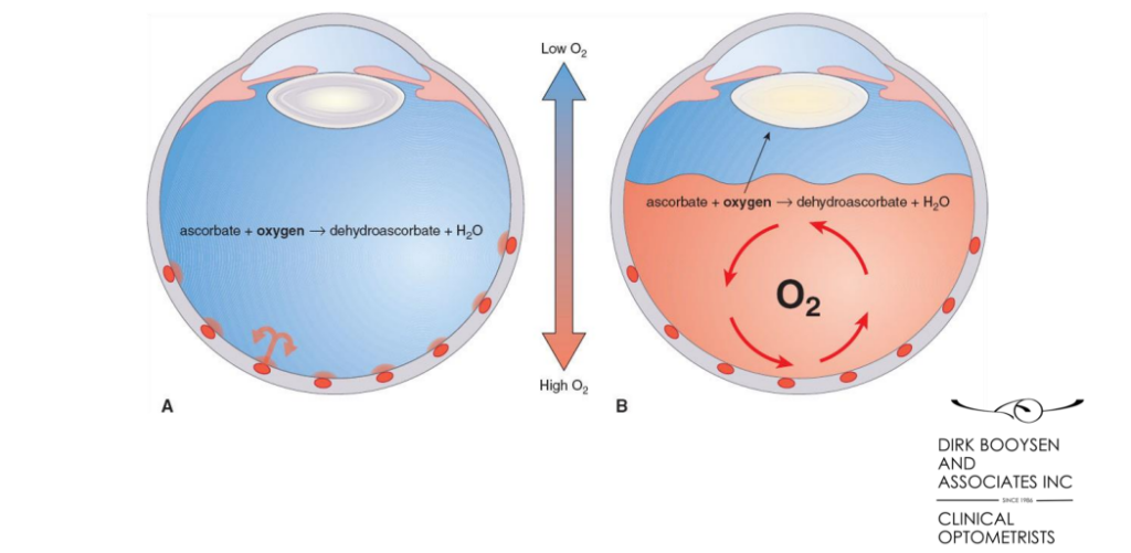
- In this regard, high levels of ascorbic acid (Vit C) may play a protective role as a scavenger of free radicals resulting from retinal and lens metabolism as well as free radicals generated by photochemical reactions
The vitreous as a physiological and metabolic buffer
Vitrectomy and cataract
Degeneration of the vitreous either by age related liquefaction (syneresis) or by it’s removal (PPV) during retinal surgery, increases the exposure of the anterior segment structures, including the lens, to higher O2 levels
The lens and trabecular meshwork, normally functioning via anaerobic metabolic pathways, are exposed to higher O2 levels, which results in oxidative damage to proteins and lipids – formation of nuclear cataracts and higher risk of glaucoma
Vitreous examination
Because of its relative transparency and low reflectivity, the vitreous is a difficult structure to study
Vitreous examination by optical means is based mainly on observation of the Tyndall phenomenon, which is caused by the scattering of light by particles in transparent media
This examination must be carried out in an optical section using a well-focused narrow slit beam and a good quality (low distortion and wide field of view) pre-corneal lens
The light should form a sharp and bright slit, and make as large an angle as possible with the slit-lamp

Vitreous examination – Mydriasis
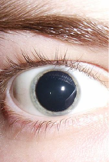
- Full mydriasis and clear ocular media are also of prime importance for adequate vitreous examination – Cyclomydril rather than Mydriacyl?
- Retro-illumination is useful in detecting particles in the vitreous cavity and on the inner retinal surface, as well as new vessels on the inner retinal surface, and minimal vitreous detachment
- To enhance the Tyndall effect and the visibility of fine details in the vitreous cavity, a strong source of blue or green monochromatic light can be used
Dynamic and static examination
- Dynamic examination is performed by asking the patient to look rapidly up, down, left or right, then back to the primary position of gaze
- These movements disturb the configuration of the vitreous body, which continues to move after the eyeball has become stationary – particles suspended in the inferior vitreous become visible
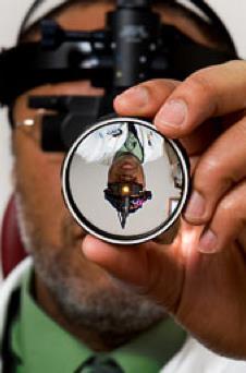
Binocular indirect ophthalmoscopy
- Image is real, inverted and laterally reversed
- Requires lots of skill to use
- Expensive – R30K without the required fundus lenses
- Options from 15D, 20D, 30D, and 40D fundus lenses which provide different levels of magnification
- Can use with scleral indentation
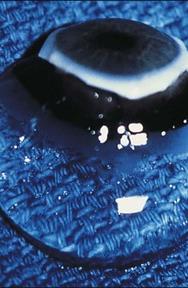
Eyesi indirect virtual reality simulator
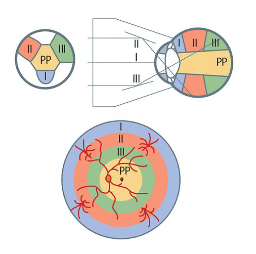
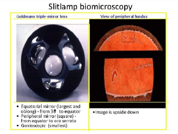
Goldmann 3 Mirror lens
- The Goldmann three-mirror contact lens neutralizes the refractive power of the cornea, allowing examination of the entire vitreous cavity
- The central portion of the lens offers a 30° upright/erect view of the posterior vitreous and of the retina
- The equatorial and peripheral mirrors are used to examine the peripheral vitreous cavity and retina from 30° to the equator and from the equatorial area to the ora serrata respectively
- The view in the mirror is inverted/upside down but not laterally reversed
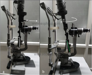
Slit lamp setup and fundus view
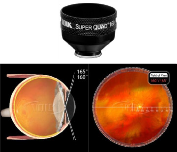
Volk Super Quad 160
- Contact lens with low magnification and large field of view
- Image is real and inverted/upside down
- Can view the entire fundus up to the ora serrata
- Relatively inexpensive – $1000
- Requires some skill to use
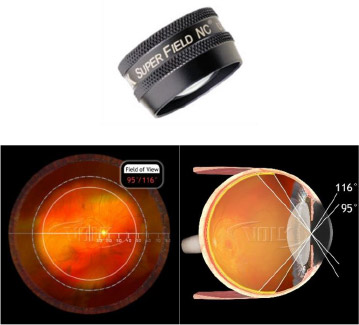
Volk Super field
- Non contact lens with relatively large field of view
- Image is real, inverted and laterally reversed
- Inexpensive – $500
- Easy to use
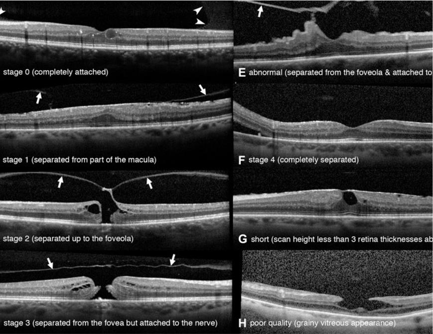
Posterior segment OCT
- Spectral-domain OCT (SD- OCT) technology allows better visualization of the posterior cortex/hyaloid/ILM/retina and a more detailed image of its structure than other technology
- Limited to the macular region and disc, little value to examine the peripheral retina
- Very expensive technology but worth the investment as it has multiple functions in practice
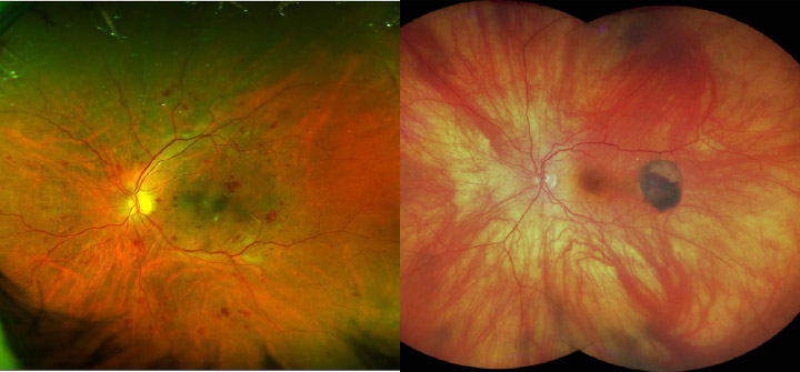
Ultra wide field fundus imaging Zeiss Clarus and Optos Daytona
A&B Scan Ophthalmic Ultrasound
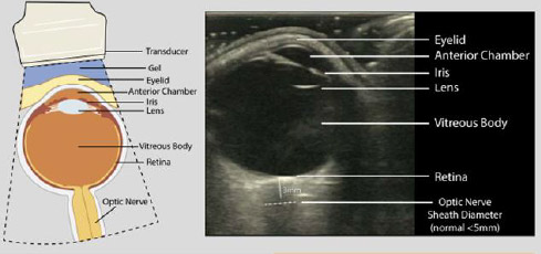
- B-scan, is a non-invasive test routinely used in clinical practice to assess the structural integrity and pathology of the eye
- Provides additional information not readily obtained by direct visualization of ocular tissues, and it is particularly useful in patients with pathology that prevents or obscures ophthalmoscopy
- A-scanning measure the length of the eye, distances within the eye, and visual axis of the eye
- The axial A-scanning was also used to help determine intraocular lens power
Symptoms and signs of troublephotopsia and phosphenes
- Extensive light flashes/photopsia forming an arc in the temporal periphery of the affected eye
- Caused by the shrunken detached vitreous that hits against the photosensitive nasal periphery of the retina when the eye moves
- Photopsia seldom occurs when the vitreous hits the temporal periphery of the retina as this retinal area is not as sensitive to light
- Photopsia must be distinguished from a sharply localized light flash, or phosphene, that is generally caused by focal vitreoretinal traction
- Sharply located phosphenes may occur in any portion of the patient’s field
- They are relatively rare, but when present, they generally manifest themselves in the lower nasal quadrant, and the vitreoretinal traction that causes them often leads to a retinal tear and detachment located in the upper temporal periphery
Other causes of photopsia and phosphenes- Wills Eye Manual
- Ophthalmic migraine – aura typically lasts 20 -30 minutes and resolves
- Pressure phosphenes due to rubbing
- Conditions that affect the function of the eye, including retinitis pigmentosa, diabetic retinopathy, ARMD, and optic neuritis (often due to MS)
- Medical issues such as brain tumours, and strokes may impair the visual regions of the brain and cause stimulation that produces a sense of seeing fleeting images of light that aren’t there
- Cerebrovascular disease (blood vessel disease in the brain) or systemic low BP (postural hypotension) can result in diminished blood supply to the brain, diminished brain function causing flashing lights or similar phenomena for a few seconds.
- Blind eye and bilateral patching
- Charles Bonnet syndrome – visual hallucinations
- Psychosis
- Parieto-temporal lesions and other CNS causes
- Medication also can alter the function of the retina in ways that produce phosphenes ivabradine, which is used to treat tachycardia (rapid heart rate) is one such drug
Schafer’s sign – types of cells found
Table 1. Types of Cells Found in the Anterior Vitreous and their Clinical
implications
| Abnormal Vitreous Cells | Source | Clinical Indication |
| Brown (Shafer’s sign) cells | Pigment from RPE of retina | Retinal break |
| Red cells | Red blood cells from haemorrhage | Retinal break or proliferative retinal process |
| White cells | Inflammatory white blood cells | Vitritis, pars planitis |
- When red/brown cells are found following an acute PVD, there’s nearly a 100% correlation with retinal tears
- If Schafer’s sign is present, the likelihood of an associated retinal tear is 52 x higher than in cases where there is absence of these signs
- 25% to 90% of retinal tears are expected to proceed to a retinal detachment, missing this warning sign could put your patient’s vision in grave danger
- White, or non-pigmented, cells in the anterior vitreous may indicate an inflammatory condition
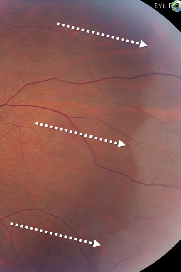
White without pressure
- Distinctive white appearance of the peripheral retina without indentation
- Whiter than the retina in white with pressure and the choroidal markings are almost obscured
- Whiteness further accentuated if scleral depression done
- Margins are sharply demarcated from normal retina
- Intervening patches of normal retina should not be confused with retinal holes
- Exact cause is unknown;
- one school of thought states it to be a manifestation of peripheral vitreous traction while one believes it to be simply an abnormal reflex from a structurally normal VR interface
- Frequently causes confusion with subclinical RD and Retinoschisis but indentation clearly reveals that retina is still apposed to the RPE
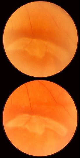
White with pressure
- Distinctive milky white or opalescent appearance of the peripheral retina that is observed in many normal eyes when examined with scleral depression
- Retina appears normal without depression
- It is common and seen in around 30 to 35% of eyes examined with scleral depression
- Inferonasal quadrant least likely to be affected
- Incidence increases with age, no sex predilection
- Benign condition not associated with retinal breaks – associated with strong vitreoretinal adhesions
- Must be carefully distinguished from a subclinical peripheral RD
Vitreous detachment – PVD
One study reported the presence of vitreous detachment in 6% of normal subjects between 45 and 65 years of age and in 65% of those between 65 and 85
The vitreous degenerates gradually by partial liquefaction – synchesis and by shrinkage of the gel – a process called syneresis
The vitreous gel loses much of its water and shrinks like a sponge from which water is expelled
The condensation of the intra vitreous fibrils causes vitreous opacities -floaters The shape of the floaters depend on the shape of the defect in the posterior hyaloid membrane where it was once attached to the optic disc – Weiss ring
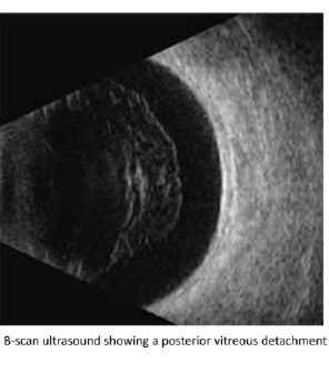
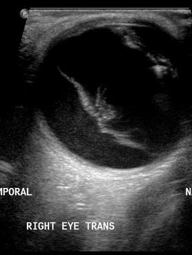
B-Scan and PVD
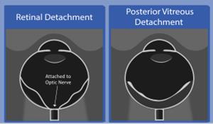
- PVD is more mobile than RD
- A-scan spike is higher in RD than PVD
- PVD is more prominent with higher gain settings while RD remains prominent with low gain settings
- PVD membrane has more uniform thickness than RD
- PVD variable attachment at the disc
- RD always attached at the disc
Vitreous changes and retinal detachment
- The close relationship that exists between vitreous changes and retinal detachment has been so solidly established over > 100 years that rhegmatogenousretinal detachment is thought of as a vitreoretinal disease
- A retinal break most frequently results from vitreous traction after a PVD has developed
- Two main factors are responsible for the development of retinal breaks and retinal detachment:
- localized vitreoretinal adhesions and traction,
- and further effects of synchesis and syneresis
Development of rhegmatogenous retinal detachment – RRD
- Fluid vitreous is indispensable to the development of retinal detachment
- Fluid vitreous must be able to reach freely the area of a retinal break in the subhyaloid space either through the anatomical holes that exist in the vitreous cortex in front of the macula and disc, or through a tear in the cortical vitreous
Development of RRD continued
- Once the retina is torn, the fluid enters into direct contact with the photoreceptors and pigment epithelium
- At this point the fluid vitreous probably contributes to detaching the retina
- The fluid vitreous weakens the chorioretinal adhesion by a double mechanism
- The first is by diluting the glycosaminoglycans that serve as an adhesive between photoreceptors and pigment epithelium
- The second mechanism probably works by overwhelming the pump action that exists in the RPE and maintains the subretinal space relatively fluid-free in a normal eye
Development of RRD – summary
- The availability of liquid vitreous is almost as important as vitreous traction in the pathogenesis of rhegmatogenous retinal detachment – RRD
- In the absence of fluid vitreous, a retinal break generally fails to be accompanied by a retinal detachment and heals spontaneously
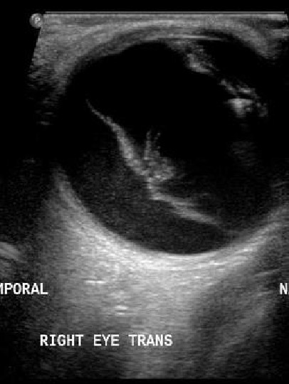
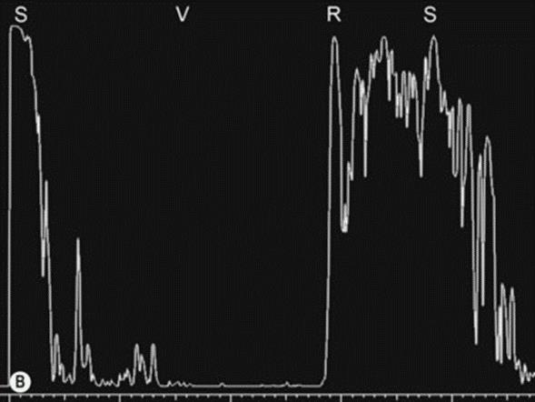
A & B-Scan of RD
- Total funnel shaped RD attached at the disc
- B-Scan with low (49dB) gain setting
- A-Scan shows 100% peak corresponding to RD S – sclera, V – vitreous, R – retina,
Other risk
- Axial myopia
- Myopic/fibrillar gel
- Early blue-green cataract
- Family history of retinal tear or detachment
- Lattice retinopathy
- Hereditary vitreoretinopathies e.g. Stickler syndrome
- Congenital ocular abnormalities e.g. glaucoma, cataract
- Retinopathy of prematurity
| Symptoms of PVD | Symptoms of retinal tear | Symptoms of retinal detachment |
| Temporal arc of light particularly noticed in dim light and associated with eye movement One or two floaters described as a ring, tadpole, fly or spider | May have symptoms of PVD Sudden shower of hundreds of dots (pigment or red blood cells) Cobweb-like ‘film’ suggests haemorrhage | ‘Shadow’ or ‘curtain’ or reduced visual field May have had preceding symptoms of PVD +/- retinal tear Loss of central vision Bright, possibly multi-coloured, lights over whole visual field |
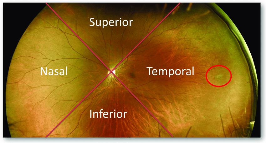
Distribution of retinal breaks in RD 60% in the upper/superior temporal quadrant 15% in upper nasal quadrant 15% in lower nasal quadrant
- 60% in the upper/superior temporal quadrant
- 15% in upper nasal quadrant
- 15% in lower nasal quadrant
- 50% of eyes with RD have more than one break – usually located within 90
degrees of each other
The upper/superior temporal quadrant is the most common site for retinal break formation and should be examined in detail if a break is not readily detected
Posterior hyaloid membrane detachment – PHMD and Vitreomacular adhesion – VMA
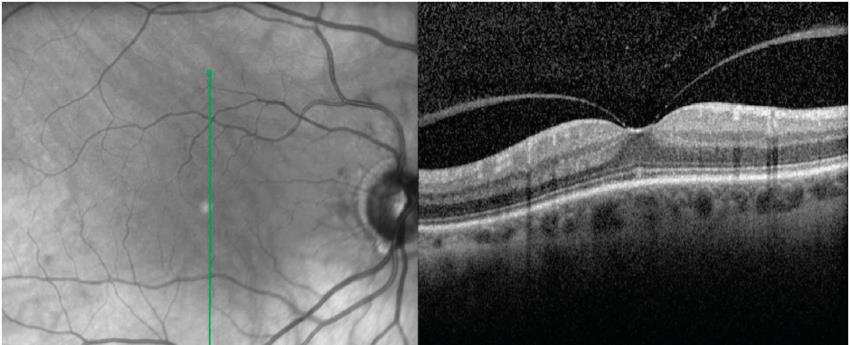
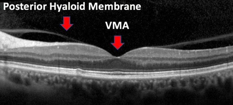
- The diagnosis of VMA is applied to patients who have incomplete separation of the posterior vitreous with persistent attachment to the macula
- In the past, VMA has been classified by symptomatic patients versus asymptomatic patients (i.e., based on a patient’s visual complaints)
- With the introduction of OCT, physicians have become aware that VMA is a more common entity than was previously clinically known and may be part of the normal formation of PVD
VMA
- Vitreomacular adhesion is typically asymptomatic and non-pathologic, and does not cause any apparent retinal changes
- It is a natural component of the development of a PVD and can therefore be considered an incomplete PVD
- Vitreomacular adhesion, although asymptomatic, has been hypothesized as playing a role in the pathogenesis of many macular conditions such as neovascular ARMD, macular hole, and diabetic macular edema
Vitreomacular traction – VMT
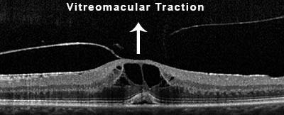
- In abnormal vitreous adhesion, there can be excessive traction on the macula from the vitreous that changes the contour of the foveal surface
- There may be a distortion of the fovea, a blunted foveal reflex, cystic changes, or (in severe cases) subretinal fluid
- OCT may be the only feature that distinguishes VMT from focal VMA when the retinal anatomy is otherwise normal
- In addition, there may be elevation of the retina at the fovea at the level of the RPE
- The combination of anatomical changes on OCT with signs of perifoveolar PVD constitutes a diagnosis of VMT
- VMT can be associated with thickening of the macula, vascular leakage on FA, macular schisis, and CME
- The anatomical changes to the fovea induced by VMT can lead to reduced BVA, metamorphopsia, and micropsia
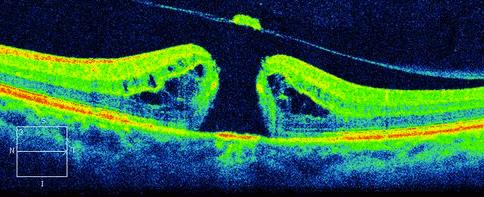
Macular hole FTMH
- A full-thickness macular hole (FTMH) is a full thickness defect in the fovea, and includes the complete interruption of all neural retinal layers from the ILM to the RPE
- Anteroposterior traction, secondary to abnormal vitreoretinal attachment at the fovea, and tangential contraction of the perifoveal vitreous cortex may be responsible for the development of macular holes
Primary and secondary macular holes
- A primary/idiopathic macular hole is caused by vitreous traction on the fovea from an abnormal vitreous separation.
- A secondary macular hole is caused by other pathologies not associated with previous VMT
- – Examples include blunt trauma, high myopia, macular telangiectasia type 2, surgical trauma, and other causes of macular oedema
Epiretinal/Preretinal membrane – ERM
- ERM is a cellular proliferation that creates a semitranslucent, fibro cellular proliferation on the surface of the inner retina
- Formed by glial cells that normally reside in the superficial retina and proliferate over the inner retinal surface
- Avascular and can form surface wrinkling of the retina – cellophane retinopathy
- Collagenous fibrils from the vitreous may be interposed between the ILM and the ERM
- ERMs are associated with retinal folding and macular thickening
- decreased BVA, metamorphopsia, micropsia, and monocular diplopia
- Epiretinal membranes affect approximately 7% of the population and are bilateral in 10-20% of patients
- In idiopathic cases of ERM, cellular proliferation may be associated with an incomplete PVD
- Epiretinal membranes can also arise secondary to retinal vascular disease, diabetes, trauma, inflammatory conditions, tumors, and retinal dystrophies
Lamellar macular hole – LMH
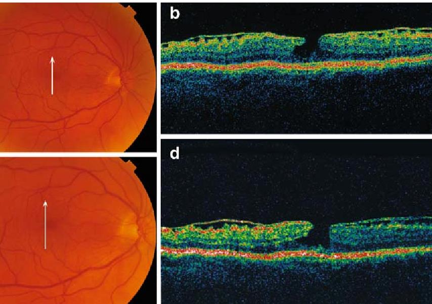
- In 1975, Gass described a lamellar hole as a macular lesion resulting from cystoid macular edema
- A lamellar macular hole (LMH) is now known as a partial thickness foveal defect; however, there is no universally accepted definition of lamellar macular holes
- Many believe they arise from incomplete full-thickness macular hole formation
- ERMs are commonly associated with eyes that have lamellar macular holes.
- The pathogenesis, configuration, and progression of lamellar macular holes are therefore believed to be affected by tangential retinal traction due to ERM contraction
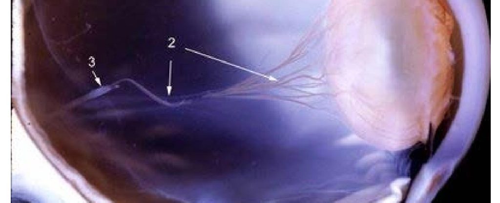
Vitreous opacities
Failure of the hyaloid system to regress
- Mittendorf’s dot
- Muscae volitantes
- Persistent hyaloid system
- Bergmeister’s papilla
Degenerative vitreous changes
- Asteroid hyalosis
- Cholesterosis bulbi/synchesis scintillans
- Amyloidosis of the vitreous
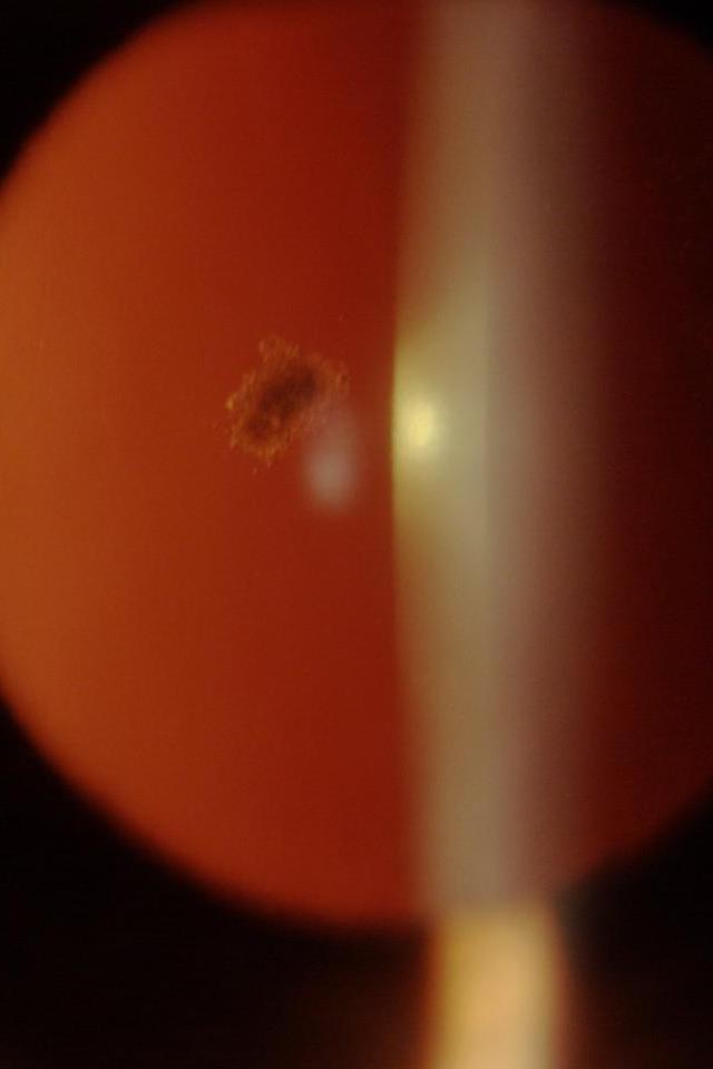
Mittendorf dot
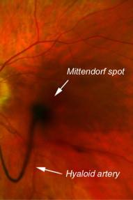
- Prominent fibro-vascular plaque on the posterior surface of the lens, slightly inferior and nasal to the visual axis
- Original site of communication of the hyaloid artery to the tunica vasculosa lentis
- Generally, it is a small dot on the posterior capsule, occasionally with a small strand of residual anterior
hyaloid attached to it Muscae Volitantes Persistence of the vasa hyaloidea propria with multiple small remnants may lead to symptoms of floaters or “muscae volitantes”
Muscae Volitantes
- Persistence of the vasa hyaloidea propria with multiple small remnants may lead to symptoms of floaters or “muscae volitantes
Persistent hyaloid system
- Persistence of only the anterior part of the hyaloid artery may occur with a residual vessel attached to the posterior surface of the lens, usually inferior and nasal to the lens center
- Persistence of the posterior portion of the hyaloid artery, which is the last part of the hyaloid system to regress during fetal development, occurs in 95% of premature infants and 3% of full term infant with a variable amount of associated fibrous or glial tissue
Bergmeister’s papilla
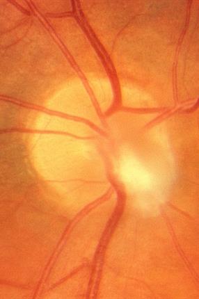
- Bergmeister’s papilla, a common embryonic remnant, may represent both deficient atrophy of glial tissue and glial overgrowth normally located at the optic disc
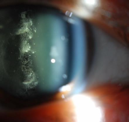
Asteroid hyalosis
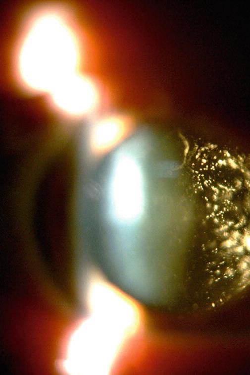
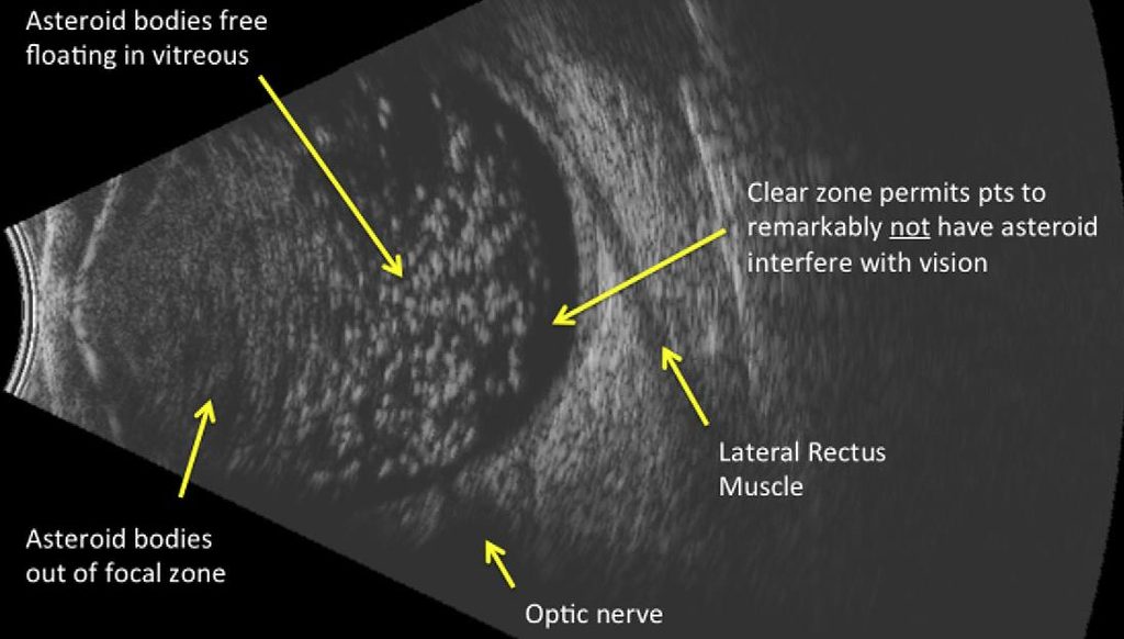
- Striking clinical appearance of glistening, yellowish white, spherical bodies suspended throughout the vitreous
- The asteroid bodies are variable in size and do not appear to be affected by gravity
- Vitreous gel shows little or no evidence of liquefaction surrounding these particles
- The patient is usually completely asymptomatic, with no decrease in visual acuity
- Complex lipid and calcium phosphorus composition
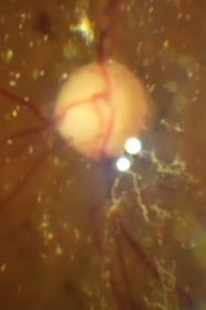
Synchesis scintillans/Cholester osis bulbi
- Synchesis scintillans crystals are cholesterol crystals
- More appropriate term for synchesis scintillans is cholesterosis bulbi
- The exact aetiology of the cholesterol crystals in cholesterosis bulbi is uncertain
Amyloidosis of the vitreous
- Amyloidosis is a disease caused by localized or generalized deposition of an abnormal protein in the body
- Opacification of the vitreous caused by amyloid is an uncommon and often misdiagnosed condition that causes progressive visual loss
- The process is usually associated with primary familial systemic amyloidosis
- Ocular involvement was found in 8% of cases of familial amyloidosis but the incidence may be higher
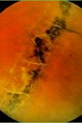
Lattice degeneration
- Progressive lattice like degeneration seems to result from insufficiency of the retinal circulation or from other factors markedly affecting the inner retinal layers and the vitreous cortex.
- The affected retina is thin and degenerated, and a vitreous condensation of variable thickness is attached to the anterior and posterior edges of the lattice
- The membrane contains vitreous fibers that seem to exert traction on the edges of the lattice like degeneration
- Lattice like degeneration affects the retina, the cortical vitreous, and the choroid
- The zone involved is especially prone to develop retinal breaks – round holes
- The condition is usually bilateral, and it is found predominantly in myopic eyes
- It is characterized by retinal thinning, variable pigmentation, and choroidal alterations
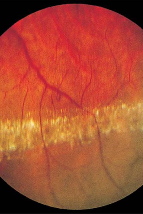
Snail track degeneration
- Snail track degeneration forms sharply demarcated bands of white, wrinkled changes of the inner retinal surface, reminiscent of tracks made by a snail
- It has been called “cellophane-like” and may also occur in the macula. It is made up of cells that form a membrane anterior to the inner retinal surface
- Vitreoretinal adhesions may form at the margins of this degeneration which may have a rhegmatogenous potential
- Round holes without opercula may also develop
- Fibrillary changes of the vitreous body may occur with syneresis and optically empty liquefied pockets
- Snail track degeneration seems to occur more often in myopic eyes Don’t forget about the vitreous!
Thank you
- Don’t forget about the vitreous!
- Flashes and floaters require careful further investigation
- Remember PVD’s relationship to RRD
- Look for horseshoe tears in the superior temporal retina
- Remember the vitreomacular relationship
- Schafer’s sign is nearly 100% correlated with retinal tears